• Ouhai, Wenzhou, Zhejiang China
  • 24/7 Service

Top-Tier Hospitals

9000+ Beds Combined

World-Renowned Doctors

Transplant Pioneers

Dedicated Care Team

Satisfied Patients
Call Us : +86 18357823575

Category Archives: Uncategorized

81-year-old patient with coronary heart disease gets a new lease on life!

81-year-old patient with coronary heart disease gets a new lease on life!

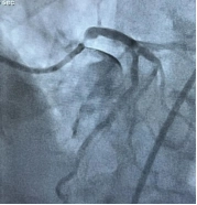
A 81-year-old patient with a history of coronary heart disease for several years was admitted to the Department of Cardiovascular Medicine. Coronary CT performed previously had indicated severe vascular calcification, and the patient had been managed with conservative medical therapy at that time. Two days before presentation, the patient experienced recurrent angina attacks. Electrocardiography showed myocardial ischemia, and myocardial enzymes were elevated. Based on the patient’s typical symptoms and coronary CT findings, the Department of Cardiovascular Medicine diagnosed acute coronary syndrome, and the patient was immediately admitted to the Cardiac Care Unit. On the day of admission, emergency coronary angiography was performed, revealing severe vascular calcification, 95% stenosis at the ostium of the left main coronary artery, and near-total occlusion of the right coronary artery. Considering the clinical history, the culprit vessel was determined to be the right coronary artery. The right coronary artery was opened with the assistance of multiple devices, and one stent was implanted. The chest pain was relieved.

5c714a41a85355abe5224cef92c5e82e6926c490338d2c3e3e080307ddc506dc

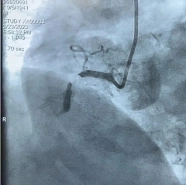
Coronary angiography findings: Near-occlusion of the right coronary artery, severe stenosis at the ostium of the left main coronary artery.

After the emergency procedure, the patient no longer experienced chest pain at rest. However, the most important coronary vessel—the left main coronary artery—still had 95% stenosis with severe calcification, raising the question of how to manage it. The Department of Cardiovascular Medicine held a discussion and proposed the following options: 1. Conservative therapy: Since the patient’s symptoms had been relieved, could medical conservative therapy be considered? However, given the severe vascular stenosis and a functional assessment QFR = 0.64 indicating severe ischemia, conservative therapy would carry a high risk of sudden death after discharge. 2. Coronary artery bypass grafting (CABG): The patient had recently suffered myocardial infarction and undergone percutaneous coronary intervention and was currently taking antiplatelet drugs, so surgical intervention would carry a high bleeding risk. In addition, the patient and family considered surgery too invasive and declined CABG. 3. Percutaneous coronary intervention (PCI): The patient had an ostial left main lesion, and intraprocedural risks included hypotension and arrhythmia. Severe vascular calcification might require coronary rotational atherectomy. PCI was recommended to be performed under strong circulatory support. After consultation among the Heart Center, ICU, and Department of Anesthesiology, and after full communication with the patient and family, it was ultimately decided to perform PCI under ECMO (extracorporeal membrane oxygenation) support.

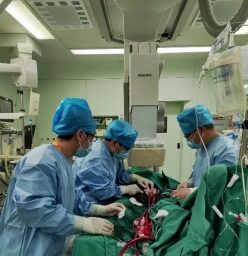
After careful planning, the procedure was performed. The Department of Anesthesiology decided to administer non-intubated general anesthesia to minimize patient discomfort. Two ICU physicians performed arteriovenous catheterization and ECMO initiation. Vascular surgeons assisted in establishing vascular access. Once everything was ready, three physicians from the coronary heart disease team of the Department of Cardiovascular Medicine performed the interventional procedure. The procedure was even more difficult than anticipated. The patient’s left coronary ostium was low, and with concomitant stenosis, multiple types and sizes of guiding catheters could not be properly engaged; ultimately, a JL4 catheter was barely engaged. Intravascular ultrasound (IVUS) revealed severe left main stenosis with nearly 300 degrees of calcification. During balloon pre-dilation, the patient’s blood pressure dropped and severe angina occurred, with blood pressure falling to as low as 60/30 mmHg. At this critical moment, the ECMO team rapidly adjusted ECMO parameters, and the cardiovascular medicine team, after rapid positioning, successfully implanted one stent. The patient’s symptoms gradually resolved, and blood pressure recovered. After multiple balloon post-dilations, intravascular ultrasound confirmed accurate stent positioning and satisfactory expansion. Final angiography showed complete resolution of the left main stenosis. ECMO was successfully weaned after the procedure, and the patient returned safely to the ward.

Procedure: ECMO initiation and percutaneous coronary intervention.

This complex coronary intervention was completed jointly by the Department of Cardiovascular Medicine, ICU, Department of Interventional Vascular Surgery, Department of Anesthesiology, and the Interventional Procedure Room, providing the patient with a safer, more minimally invasive, and more comfortable treatment strategy. This demonstrates the advantages of multidisciplinary team (MDT) diagnosis and treatment in managing complex and critically ill cases and will bring benefits to more similar patients.

What is ECMO?

ECMO stands for extracorporeal membrane oxygenation, also known as an artificial heart-lung machine. It works by draining blood from the body through blood vessels into an extracorporeal heart-lung machine for pressurization and oxygenation, and then pumping it back into the body. It can provide temporary cardiopulmonary support for patients with severe cardiopulmonary failure, buying precious time for the rescue of critically ill patients. This device became widely known to the public over the past 3 years and saved many patients with severe COVID-19. It is currently mainly indicated for patients with cardiac arrest, acute severe heart failure, and acute severe respiratory failure.

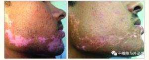
Intravenous infusion of cell therapy is a safe and effective treatment for vitiligo.

Vitiligo is an acquired, chronic, and recurrent skin disorder characterized by patchy depigmentation. It is caused by the destruction of epidermal melanocytes.

Vitiligo can be disfiguring, often leading to varying degrees of psychosocial distress and a decline in patients’ quality of life. Currently, the etiology of vitiligo has not been fully elucidated, and effective treatment options remain limited. Therefore, there is an urgent need to find an effective therapeutic approach for vitiligo.

Research on the pathogenesis of vitiligo indicates that immune mechanisms, along with other pathogenic factors, play a crucial role in the onset and progression of the disease; this significance has been confirmed by numerous studies.

1701510163554841

1. The Clinical Challenge: Up to 80% of Patients Lack Effective Treatment

Clinical data reveals that 80% of vitiligo patients do not receive effective treatment, and the ineffective rate of various therapies for vitiligo patients is as high as 50%.

While vitiligo does not affect the body’s physical health or physiological functions, its visual impact places a tremendous emotional and mental burden on patients.

2. The Breakthrough: Unique Advantages of Mesenchymal Stem Cells (MSCs)

Mesenchymal Stem Cells (MSCs) have garnered significant attention due to their unique biological properties, including:

  • Immunomodulatory properties

  • Promotion of tissue regeneration

  • Low post-transplantation rejection rates

Latest Clinical Evidence:

A recent study in India involving cell transplantation therapy for vitiligo patients showed that transplantation efficiency can be predicted by the infiltration of CD8+ T cell activity around the lesions. Meanwhile, the results demonstrated that these cells could significantly inhibit the skin-homing activity of CD8+ T lymphocytes, suggesting the great potential of cell therapy in improving the transplantation efficiency and efficacy for vitiligo.1701510194945832

3. Restoring Skin Tone: Clinical Practice of Stem Cell Therapy

Clinical researchers at Noble Clinic in Bangalore, India, pointed out that with recent advances in biotechnology and the emergence of new technologies such as cell transplantation surgery and stem cell therapy, it is now possible for the majority of patients to restore their skin tone.

Researchers explained that stem cells are injected intravenously into the body, which helps stabilize the spread of vitiligo and promotes pigmentation over time. This is because these cells possess superior immunosuppressive and immunomodulatory properties.

Most vitiligo patients suffer from autoimmune diseases, in which the patient’s immune system malfunctions and produces autoantibodies—proteins that destroy color-producing cells in the skin called melanocytes. Once these cells are killed, pigmentation does not occur, and the skin turns white. The intervention of stem cell therapy precisely blocks this destructive process from its immunological root.

4. Policy Empowerment: Accelerating Innovation in the “Lecheng Medical Pilot Zone”

As a “special zone” for medical innovation in China, the Boao Lecheng International Medical Tourism Pilot Zone enjoys core policy advantages such as “special licensed medical treatment, special research, and special operation.” Here, cutting-edge technologies like mesenchymal stem cell therapy can leverage the pilot policies granted by the state to accelerate clinical translation and application. This allows domestic patients to access internationally advanced cell therapy technologies without traveling abroad, bringing true “new hope” to the clinical treatment of vitiligo.

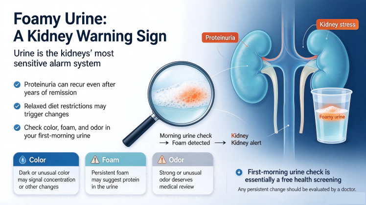
Foamy Urine: A Kidney Warning Sign

Foamy Urine: A Kidney Warning Sign

Foamy Urine: A Kidney Warning Sign

“How did you find out?” Dr. Wang Tiemin, the expert and hospital director, asked the patient and family members before him.

Outside, the rain was pouring relentlessly, stirring up veil after veil of mist. In the consultation room sat a returning patient. After several years without a follow-up visit since recovery, the once slightly soft and youthful face had grown angular and defined.

When the patient first came to the hospital in early 2018, he was tangled in worry and distress over “urinary protein.” After systematic treatment integrating Chinese and Western medicine, his urinary protein turned negative, and 24-hour urinary protein dropped to the normal range. He continued taking herbal medicine for consolidation, and follow-up reviews kept the condition stable.

Meanwhile, the weight of the word “kidney disease” gradually faded. At first, it was a boulder lodged in the hearts of the patient and his family; as the treatment took full effect, it was worn down into pebbles, then gravel, until it became a weightless speck of dust — no weight in the heart, invisible to the eye. When the suffering lost its weight and its shape, everything returned to how it was before.

After Stabilization, Dietary Restrictions Were Fully Loosened

As the condition stabilized, the patient’s diet opened up completely. According to what was learned, high-salt, high-sugar, and high-fat meals, ice-filled coffee and cold drinks, beef and mutton that easily provoke the immune system — foods once strictly avoided — had become ordinary daily fare. The kidney-damaging components and metabolic burdens in these meals accumulated in the body once again, slowly overwhelming the metabolic balance restored by medication.

“Just the past couple of days I saw foam in his urine. I said, no, we need to get this checked.” The patient’s father was the first to notice the abnormality.

Foamy Urine, in Medical Terms, Is “Proteinuria”

If the everyday phenomenon of “foam in urine” is translated into medical terminology, it is “proteinuria.” Protein leaking from the kidneys changes the surface tension of urine — much like adding egg white or dish soap to water — causing large amounts of foam that does not easily dissipate. This is an abnormal signal mentioned by many patients at their first visit, and it covers a very wide range of conditions.

There are many types of kidney disease — IgA nephropathy, membranous nephropathy, hypertensive nephropathy. Why is proteinuria the first to appear in all of them?

Because what is most fragile is always the first to give way. For the kidney, the most fragile part is the filtration barrier. It blocks proteins based on molecular size and charge; structurally, it is like an extremely “fine sieve.”

Because it is so fine, any slight damage from inflammatory reactions or metabolic attack will impair the structure of the filtration barrier. In primary nephritides such as IgA nephropathy and membranous nephropathy, the deposition of immune complexes directly damages the basement membrane or podocytes, causing “holes in the sieve” of the filtration barrier and allowing protein to leak out. Likewise, metabolic attack from high glucose toxicity directly injures podocytes — which can barely regenerate — and once damaged, the negative charge barrier on the “sieve” disappears, also leading to protein leakage.

Therefore, proteinuria is the earliest and most sensitive marker of glomerular injury, and foamy urine has become the most common kidney disease signal.

Urine Self-Check: A Free Checkup

This also shows how keenly urine reflects abnormalities in the body. From a health-warning perspective, a self-check of urine is equivalent to a free checkup. Morning urine in particular — stored in the bladder for 6–8 hours, undisturbed by food and activity, and concentrated overnight — can amplify abnormal signals to the greatest extent.

It is recommended to self-check from the following 3 aspects:

1. Look at Color

  • Normal: pale yellow or amber.

  • Dark tea-colored or soy-sauce-colored: be alert to liver/gallbladder problems or muscle breakdown.

  • Meat-washing-water-colored or red: may indicate stones, infection, or kidney problems.

  • Rice-water-like milky white: watch for lymphatic or urinary tract infection.

2. Look at Foam

If the foam is fine and dense and does not dissipate after ten-plus minutes, it suggests urinary protein may be elevated — a typical sign of kidney injury.

3. Smell the Odor

  • Slight ammonia odor: normal.

  • Rotting-apple odor: be alert to diabetic ketoacidosis.

  • Foul odor: may indicate severe urinary tract infection.

When to Seek Medical Attention

If the above abnormalities persist for more than 3 days, or are accompanied by edema, fatigue, or lower back pain, please seek medical attention promptly for a urinalysis + kidney function test.

If it is only occasional dark color or strong odor, it is most likely due to drinking too little water or staying up late — drink two more glasses of water today, check again tomorrow, and there is no need for excessive anxiety.

The premise of a free checkup is proactive awareness. Only when you know the “signals” in your heart can you match them to the right “seat” — or rule them out directly.

Headache

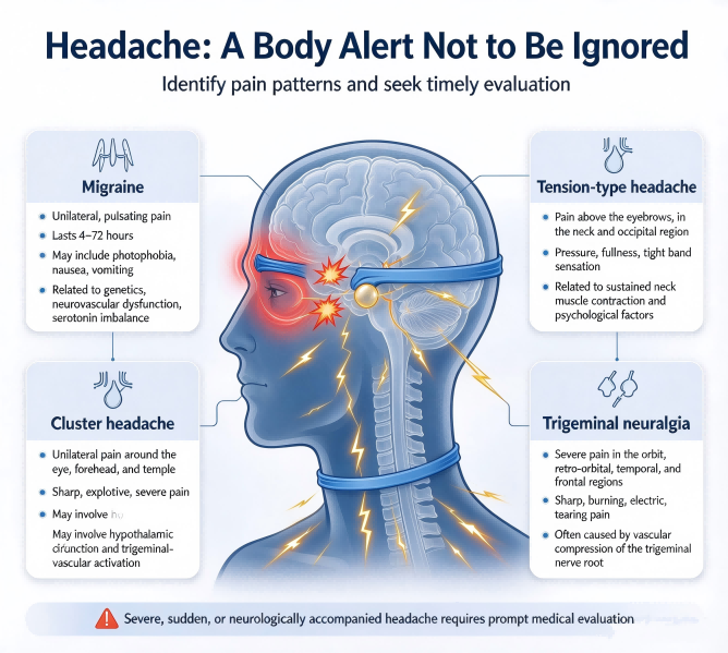
Headache: A Body Alarm That Should Not Be Ignored

Headache: A Body Alarm That Should Not Be Ignored

Nearly everyone has experienced a headache at some point, but many people dismiss it—assuming it will pass on its own or that “a painkiller will do.” In fact, severe headaches can be a warning sign of serious illness, and enduring them or self-medicating may delay treatment.

The following introduces common conditions that present primarily with headache.


I. Primary Headaches: The “Top Troublemaker” of Chronic Recurring Episodes

Primary headaches are those with no clear organic lesion or systemic disease causing them.

1. Migraine

Prevalence: Lifetime prevalence of about 18%, more common in women than men.

Features:

  • Often occurs on one side of the head, with unilateral, pulsating or throbbing characteristics

  • Typically lasts 4–72 hours, and may be accompanied by photophobia, nausea, vomiting, and other discomfort

  • Some patients experience blurred vision and other symptoms, which can severely affect daily work and life

Causes: Related to genetics, neurovascular dysfunction (such as activation of the trigeminovascular system), and imbalance of brain neurotransmitters (such as serotonin).

2. Tension-Type Headache

Incidence: The most common headache type in adults, slightly more common in women.

Features:

  • Slow onset, gradually increasing in severity, and may last for days or years

  • Pain is located above both brow ridges, in the neck, and in both occipital regions, often appearing after emotional stress, excitement, fatigue, or staying up late

  • Often presents as dull pain, like wearing a hat on the head, with a sense of pressure, fullness, tightness, or band-like constriction; may also present as heaviness in the head and shoulders

Causes: Sustained contraction of head and neck muscles (such as prolonged tension or strain) stimulates nerve endings within the muscles to cause pain; psychological factors (such as anxiety and depression) may also play a role.

3. Cluster Headache

Rarity: Male incidence is about 5–6 times that of females, with onset mostly between ages 20 and 40.

Features:

  • Mostly acute-onset severe unilateral headache, with recurrent episodes

  • Pain is mostly located in the unilateral orbit, supraorbital area, retrobulbar area, and temple

  • Presents as sharp, explosive, non-pulsating severe or extremely severe pain

Causes: May be related to abnormal hypothalamic circadian function, activation of the trigeminovascular system, histamine release, or genetic factors; the specific mechanism remains unclear.

4. Trigeminal Neuralgia

Predominant Population: Middle-aged and elderly people aged 48–59, slightly more common in women.

Features:

  • The most intense pain is concentrated in the orbit, retrobulbar area, temporal region, and frontal region, sometimes involving the occipital region, maxilla, neck, ear, and teeth, and occasionally radiating to the ipsilateral shoulder and arm

  • The pain is extremely severe, often presenting as stabbing, burning, throbbing, sharp, electric-shock-like, tearing, or drilling pain

Causes: Most cases involve vascular compression of the trigeminal nerve root (such as displacement of the superior cerebellar artery); a minority are caused by tumors, inflammation, or demyelinating lesions of the nerve.


II. Secondary Headaches: Caused by Identifiable Diseases

1. Headaches Caused by Intracranial Lesions

① Subarachnoid Hemorrhage

A very serious, sudden-onset severe headache, partly caused by increased intracranial pressure, presenting as localized or generalized headache, and may be accompanied by other central nervous system symptoms or signs, such as severe nausea, vomiting, impaired consciousness, and meningeal irritation signs (such as neck stiffness).

② Intracerebral Hemorrhage

Sudden severe headache, vomiting, impaired consciousness, and focal neurological deficits (such as hemiplegia and aphasia), mostly caused by vascular rupture due to hypertension or vascular disease; CT can quickly confirm the diagnosis. The condition is dangerous and progresses rapidly, requiring emergency reduction of intracranial pressure and blood pressure control; some cases require surgical removal of the hematoma to save life.

③ Cerebral Venous Sinus Thrombosis

A rare cerebrovascular disease, mainly presenting with headache, papilledema, and focal neurological deficits, often caused by increased intracranial pressure due to impaired venous return. Causes are diverse, including infection, hypercoagulable states, and trauma. Imaging examinations (such as MR venography) are key to diagnosis.

④ Intracranial Space-Occupying Lesions

  • Headache features: Sudden severe headache (peaking within seconds), lasting from minutes to hours, most obvious in the morning, may be aggravated by coughing, sneezing, and other actions, mostly located in the bilateral frontal region

  • Accompanying symptoms: Often accompanied by nausea and vomiting; altered consciousness or falls may occur

⑤ Reversible Cerebral Vasoconstriction Syndrome

Often triggered by exercise, emotional stress, coughing, etc., presenting as sudden “thunderclap” severe pain lasting from minutes to hours, which may recur and gradually worsen. About 20% of patients develop stroke (ischemic or hemorrhagic), with the postpartum period being a high-risk stage; in most patients, cerebral vasospasm resolves spontaneously within weeks without sequelae.

2. Headaches Caused by Systemic Diseases

① Hypertensive Headache

  • When intracranial vascular pressure rises sharply, headache, dizziness, nausea, and other symptoms may occur

  • Pain is mostly concentrated in the occipital and temporal regions; sometimes the head feels heavy with a sense of pressure

  • After blood pressure stabilizes, headache symptoms usually gradually disappear

② Low Intracranial Pressure Headache

  • Often moderate bilateral symmetric dull or distending pain, characterized by being obvious in sitting or standing positions and quickly relieved in the lying position

  • Patients may have nausea, vomiting, neck pain, dizziness, diplopia, blurred vision, and other manifestations; severe cases may develop cerebellar tonsillar herniation, subdural or epidural hemorrhage, and other conditions

3. Cervicogenic Headache

Pain Characteristics:

  • Clear location: Unilateral occipital or frontotemporal dull pain (occasionally bilateral), often radiating to the vertex and orbit

  • Triggering mechanism: Neck movement (such as rotation and flexion) and prolonged fixed posture (such as desk work) can aggravate the pain

  • Accompanying symptoms: Neck stiffness and shoulder-back soreness; some patients have dizziness, photophobia, or tinnitus (easily misdiagnosed as migraine)

Physical Examination and Diagnostic Markers:

  • Cervical tenderness: Obvious tenderness at the C1-C3 paravertebral area or the exit of the occipital nerve

  • Nerve block test: Headache relief after diagnostic cervical nerve block is one of the gold standards for diagnosis

Causes:

  • Cervical lesions: Disc herniation and joint degeneration (commonly at C1-C3)

  • Nerve compression: Greater/lesser occipital nerves compressed by muscle spasm or fibrous tissue

  • Secondary factors: Trauma (such as whiplash injury), long-term poor posture, or inflammation


III. Other Types of Headache

1. Sinus Headache

Features:

  • Headache is mostly dull or distending pain, accompanied by nasal congestion, rhinorrhea, and facial tenderness (such as in the forehead, around the orbit, or at the cheekbone)

  • Pain may worsen when bending over, coughing, or straining

  • Headache may be more obvious in the morning and gradually lessens over time

Causes: Inflammation of the sinuses (such as the frontal and maxillary sinuses) due to infection (viral or bacterial), allergies, or nasal polyps leads to retention of sinus secretions and increased pressure, stimulating nerves and causing headache.

2. Headache Caused by Otitis Media

Features:

  • Headache is often located around the affected ear or in the temporal region, presenting as continuous distending or throbbing pain, and may be accompanied by ear pain, tinnitus, and hearing loss

  • Pain may worsen when pressing the tragus or pulling the auricle

Causes: Middle ear infection (such as bacterial or viral infection) leads to middle ear effusion and increased pressure; inflammation stimulates ear nerves and spreads to the head, causing headache. It commonly occurs after upper respiratory tract infections such as colds and sinusitis.

3. Headache Caused by Glaucoma

Features:

  • Headache is mostly severe distending pain around the orbit and in the frontal region, and may be accompanied by nausea, vomiting, blurred vision, and halos (colored rings around lights)

  • Unilateral or bilateral episodes, easily induced at night or in dark environments

Causes: In glaucoma patients, impaired aqueous humor circulation leads to increased intraocular pressure; the high pressure stimulates intraocular nerves and surrounding tissues and is referred to the head, causing headache.

4. Medication Overuse Headache

Features:

  • Headache is chronic, bilateral mild-to-moderate distending pain or tightness, occurring daily or frequently; the original headache may be temporarily relieved by medication but worsens after discontinuation (rebound headache)

Causes: Long-term excessive use of analgesic drugs reduces central nervous system sensitivity to the drugs, paradoxically inducing or aggravating headache and forming a vicious cycle.

5. Postherpetic Neuralgia

Features:

  • Headache presents as unilateral paroxysmal stabbing, burning, or electric-shock-like pain, located in the area of previous herpes zoster rash (such as the head, face, forehead, and scalp)

  • Pain may last for months to years and recur after the rash has healed; touching the skin or mild stimulation can trigger it

Causes: After varicella-zoster virus infection, the virus latently resides in nerve ganglia; upon reactivation, it damages nerves, leading to abnormal nerve function and causing persistent neuropathic pain.


IV. Which Headaches Require Immediate Medical Attention?

  • Sudden severe headache with vomiting, confusion, or limb weakness

  • Headache accompanied by high fever and neck stiffness (beware of intracranial infection)

  • Sudden vision loss, speech impairment, or unsteady gait

  • Headache noticeably worsened by coughing or straining (beware of increased intracranial pressure)

  • New persistent headache after age 50

  • Sudden headache in pregnant or postpartum women


Knee Joint Treatment

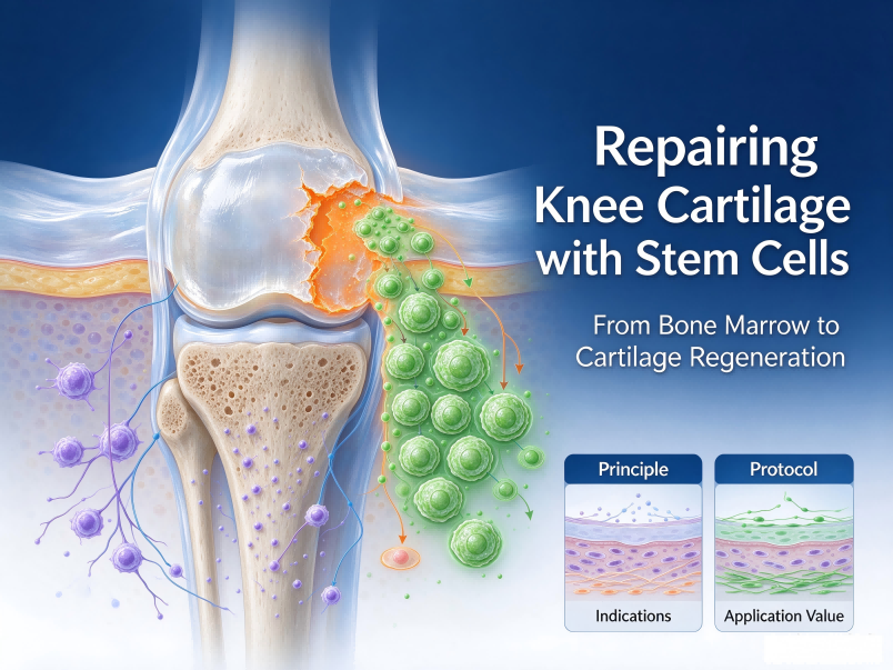
Knee Joint Injuries: How Can Stem Cell Repair Help Cartilage “Experience a Rebirth”?

Knee Cartilage Repair: How Stem Cells Bring “New Life” to Damaged Joints

Introduction: The knee is one of the most weight-bearing and frequently used joints in the human body. Once injured, it often brings long-term pain and limited mobility. Traditional repair methods are slow to recover from and have limited efficacy. However, with the development of regenerative medicine, stem cell therapy is bringing new hope to patients with knee injuries.


How Stem Cells Repair Damaged Knees

Bone marrow stem cells possess unique self-replication and regenerative capabilities, allowing them to play a reparative role in damaged areas. Specifically, bone marrow stem cells can rapidly enhance local immunity, help clear necrotic and damaged cells, and then rely on their innate regenerative ability to repair damaged knee tissues. This process regulates the local immune balance of the knee, gradually restoring the injured joint to a healthy state.

The key to this process lies in “precision.” Through precise injections, stem cells are directed to focus on repairing the affected area. This maximizes the concentration of repair power at the site of injury and enhances the efficiency of the treatment.


Customized Treatment Protocol

Stem cell therapy for knee repair typically follows a customized personal treatment plan. The general process is as follows:

  • Professional Medical Consultation: Before treatment, a professional physician evaluates the current condition of the knee injury and arranges a personalized treatment plan accordingly.

  • First Three Months (Unilateral Knee Injections): Administer one injection per month, precisely injecting stem cells into the affected knee.

  • Following Three Months (Contralateral Knee Injections): Continue with one injection per month, applying the same precise injections to the other knee.

The entire treatment course lasts for half a year. Through continuous, phased stem cell injections, it helps to gradually repair damaged cartilage and improve the overall function of the knee.


Conclusion

Knee injuries no longer have to rely solely on traditional surgery and prolonged rest. Stem cell therapy, based on the body’s own regenerative capabilities and combined with precise injection techniques and customized treatment plans, offers a new path for repairing knee cartilage damage. For those suffering from knee problems, this is undoubtedly a rehabilitation direction worth understanding and exploring.

Laryngopharyngeal cancer

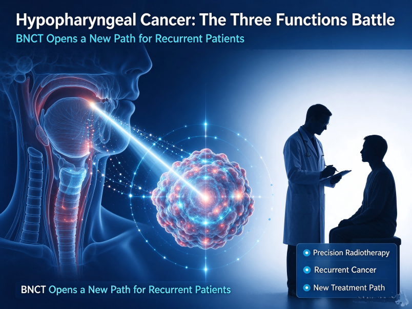
A “Three-Function Defense” in Hypopharyngeal Cancer: BNCT Opens a New Path for Recurrent Patients

The throat (pharyngolarynx) serves as a critical junction for three vital functions: breathing, swallowing, and vocalization. Anatomically, it is a confined space situated in close proximity to the carotid arteries, the thyroid gland, and the entrance to the esophagus. Hypopharyngeal cancer—most commonly squamous cell carcinoma—is often insidious; symptoms frequently appear late, meaning that by the time of diagnosis, the tumor has often already invaded surrounding structures. Among head and neck cancers, it carries a poor prognosis and a high risk of functional impairment. For a long time, the central dilemma in treating hypopharyngeal cancer has remained acute: how to achieve a radical cure while preserving the essential, yet seemingly mundane, functions of speaking, eating, and breathing?

Within standardized clinical practice, early-stage hypopharyngeal cancer can be effectively controlled through surgery or radical radiotherapy, with a reasonable chance of preserving function. However, once the disease reaches a locally advanced stage—or recurs following initial surgery or radiotherapy—treatment options become severely constrained.

Balancing radical tumor eradication with functional preservation is exceptionally difficult. Total laryngectomy or total pharyngolaryngectomy can ensure clear surgical margins, but at the cost of the permanent loss of natural speech, leaving the patient dependent on a permanent neck stoma for breathing. Furthermore, there is virtually no room for re-irradiation; the mucosal lining and cartilage of the throat have a limited tolerance for radiation, and a second course of conventional radiotherapy carries a significantly higher risk of complications, such as cartilage necrosis or severe stricture. Meanwhile, tumor progression poses a direct threat to the body’s vital pathways: airway obstruction leads to respiratory distress, while impaired swallowing prevents food intake. The condition can deteriorate rapidly, leading to a swift collapse of the patient’s overall physical state.

Boron Neutron Capture Therapy (BNCT) offers a novel approach to targeting tumors located in these “functionally critical” zones, thanks to its unique binary, targeted mechanism. In China, BNCT is currently approved for the treatment of unresectable, locally advanced, or recurrent head and neck malignancies. Unlike conventional radiotherapy, BNCT enables the precise, localized destruction of tumor cells. A boron-containing agent is administered intravenously, accumulating specifically within the tumor cells while leaving healthy tissue largely unaffected. Subsequently, the area is exposed to a neutron beam, which triggers a targeted nuclear reaction exclusively within the cancer cells. The range of the destructive particles produced by BNCT is limited to approximately the diameter of a single cell, with energy release confined entirely within the lesion. This allows for the protection of critical structures—such as laryngeal cartilage, the recurrent laryngeal nerve, the esophageal mucosa, and adjacent major blood vessels. In real-world clinical practice in China, patients with refractory laryngeal tumors—who had experienced recurrences despite multiple prior surgeries, radiation therapy, or chemotherapy—have shown significant, visible shrinkage of their lesions just 40 hours after BNCT, accompanied by a marked reduction in the sensation of a foreign object in the throat. Follow-up examinations two weeks later revealed further regression of the lesions and improved patency of the laryngeal cavity (based on individual case records; outcomes may vary).

BNCT is subject to strict clinical criteria. For early-stage hypopharyngeal cancer that can be completely resected or treated with curative-intent radiation, standard treatment protocols remain the preferred choice. Patients with unresectable tumors or late-stage recurrences should consult with specialists at an oncology hospital as soon as possible to determine their eligibility for BNCT. Candidates for BNCT must undergo a comprehensive multidisciplinary evaluation—incorporating pathological classification, imaging-based staging, and a review of their prior treatment history—before a tailored treatment plan can be established.

Advancements in cancer treatment are intended to fill gaps in conventional medicine, not to overturn existing diagnostic and therapeutic systems. For patients with recurrent or refractory hypopharyngeal cancer who have exhausted conventional treatment options, BNCT offers a new path forward: utilizing precision technology to combat stubborn disease, while employing a minimally invasive approach to preserve the vital functions of breathing, swallowing, and speech. Individuals with a long history of smoking or alcohol consumption, or those experiencing hoarseness that persists for more than two weeks, should undergo a laryngoscopic examination promptly. Patients who have already experienced a recurrence—or who have been told that “no further treatment is possible”—should avoid a “wait-and-see” approach; instead, they should seek a formal, professional evaluation, as every potential window for treatment is precious.

Jiahui Completes First International CAR-T Treatment

First Foreign CAR-T Patient Discharged in China

Since CAR-T therapy was approved for marketing in China, the Shanghai Jiahui International Cancer Center has worked with domestic and international partners to actively build an international CAR-T diagnosis and treatment platform in areas such as clinical experience sharing, academic exchange, and blood cancer education. At the same time, the multidisciplinary treatment team at Jiahui International Cancer Center has kept pace with international advanced standards and carried out a series of explorations in the clinical practice of new concepts and new technologies, hoping to bring new hope of cure to more patients with hematologic malignancies. The first foreign patient in China to receive axicabtagene ciloleucel CAR-T therapy was discharged smoothly three weeks after CAR-T cell infusion. On the day of discharge, the Jiahui medical and nursing team, who had accompanied the patient through every difficult moment of treatment, gathered with the patient’s family and friends outside the ward of the cancer center and prepared a special “discharge commemorative ceremony” for the patient to congratulate her on successfully completing CAR-T therapy.

A Foreign Mother of Three Diagnosed in Shanghai with “Primary Refractory” Lymphoma

Lyn comes from Singapore, is 40 years old, is a mother of three, and has lived in Shanghai for 15 years. Leading a healthy life, Lyn enjoys gathering with friends and has always maintained a fitness habit. In mid-2022, Lyn suddenly felt severe pain in the shoulder, neck, and upper limbs and could not sleep well all night. At first, Lyn thought it was a stiff neck, but after some time the symptoms became increasingly severe and even affected breathing. Lyn went to the emergency department of Shanghai Jiahui International Hospital for examination, where a huge mass 15 cm in diameter was found in the mediastinum. The compression from the large mass not only caused limb pain but also affected the nearby heart, great vessels, and lungs, leading to superior vena cava syndrome (SVCS), at one point causing difficulty breathing and posing an immediate life threat. The Jiahui hematology-oncology team quickly confirmed the diagnosis of “primary mediastinal large B-cell lymphoma” through biopsy pathology. This is a relatively rare lymphoma subtype, occurring mostly in young people, especially women.

After rapid confirmation of the diagnosis, the Jiahui hematology-oncology team immediately conducted an MDT multidisciplinary discussion and quickly formulated a comprehensive intensified treatment plan for Lyn. Lyn subsequently underwent comprehensive treatments including chemotherapy combined with targeted therapy and consolidation radiotherapy. Unfortunately, Lyn was insensitive to both chemotherapy and radiotherapy; the lymphoma shrank by half and then stopped shrinking, and soon began to progress. Lyn’s condition belonged to “primary refractory” lymphoma; patients who fail to achieve complete remission through standard chemoradiotherapy or who relapse and progress soon after treatment generally have a poor prognosis. Therefore, after full evaluation by the Jiahui hematology-oncology team and in accordance with international and domestic guideline recommendations, the team unanimously concluded that Lyn urgently needed more effective new treatment. The CDE website announced that CAR-T cell immunotherapy was officially approved in China for second-line treatment of adult large B-cell lymphoma, once again igniting Lyn’s hope.

CAR-T therapy, namely “chimeric antigen receptor T-cell therapy,” is a major breakthrough in individualized cellular immunotherapy for human cancer. It mainly involves collecting the patient’s T cells, performing in vitro genetic engineering modification to enhance the ability of T cells to specifically recognize tumors; subsequently, a large number of expanded “armed” T cells (i.e., CAR-T cells) are infused back into the patient to precisely kill tumor cells, and they can also form immune memory T cells, thereby achieving specific and durable antitumor effects.

Currently, the approved indications for CAR-T in China are lymphoma and myeloma. Because different patients have individualized particularities, and CAR-T therapy involves multiple key steps such as patient screening, bridging therapy, adverse reaction management, and long-term follow-up, this therapy places extremely high demands on the professionalism of the hematology-oncology team and the comprehensive diagnosis and treatment capabilities of multiple departments. Therefore, it needs to be carried out in medical centers with rich experience in hematologic tumor diagnosis and treatment and with multidisciplinary comprehensive construction.

Should they choose Shanghai, return to Singapore, or go to the United States for treatment? After careful consideration, Lyn’s family ultimately decided to undergo this internationally advanced medical therapy at Shanghai Jiahui International Hospital. Lyn’s husband shared that there were multiple factors behind this decision: first, considering the children’s schooling in Shanghai; second, the cost of CAR-T treatment abroad is far higher than in China and the waiting period is at least 3–6 months; third and most importantly, their trust in the clinical comprehensive strength and comprehensive medical support of Shanghai Jiahui International Hospital. From the first visit to the emergency department, the professionalism and meticulous care of the Jiahui team left a deep impression on Lyn’s family, and staying at Jiahui allowed them to enjoy an internationally advanced diagnosis and treatment experience in a familiar city. Facts proved that the decision at that time was very correct. The Jiahui International Cancer Center quickly launched the CAR-T clinical process, hoping to seize the best treatment timing for Lyn.

CAR-T Initiation: Everything Advanced Intensely but Orderly

As the first international hospital in Shanghai able to carry out CAR-T therapy, Shanghai Jiahui International Hospital attached great importance to this project. For this treatment, departments including the cancer center, cardiovascular medicine, neurology, ICU, respiratory medicine, gastroenterology, nephrology, imaging, laboratory medicine, and pharmacy, as well as the cancer center nursing team, collaborated closely, repeatedly conducting multiple rounds of training and scenario simulations in the early stage and promptly updating the patient’s condition.

After preliminary bridging therapy, infection screening, and pre-collection confirmation, the Jiahui hematology team collected Lyn’s cells and successfully completed the infusion of CAR-T cells.

After completion of the infusion, the Jiahui multidisciplinary team stood ready and closely monitored changes in Lyn’s various physical indicators and symptoms. On the 7th day after infusion, Lyn developed CRS symptoms such as fever, hypotension, and vomiting. The Jiahui medical and nursing team promptly carried out treatment according to the emergency plan previously formulated, bringing the condition under control in a timely manner. After CAR-T cell infusion, the “T-cell special forces” entering the body expanded well, and the patient’s pain symptoms caused by the tumor were relieved. After clinical evaluation confirmed stable condition, Lyn smoothly “exited the unit,” returning from the separately set CAR-T dedicated isolation ward to the general oncology ward. Lyn was discharged smoothly. Although long-term monitoring and subsequent treatment were still needed afterward, the most difficult hurdle had been safely passed.

Cutting-Edge Therapy Lands in China, Multidisciplinary Professional Team Provides Support

For Lyn, Shanghai is a city of different significance. During the 15 years Lyn and family lived in Shanghai, they not only witnessed the gradual improvement of Shanghai’s overall international image but also personally experienced that the application of new medical technologies in Shanghai is advancing with great strides toward the global frontier level of Europe and the United States. It is understood that Lyn was the first CAR-T patient at Shanghai Jiahui International Hospital and also the first foreign patient in China to complete axicabtagene ciloleucel CAR-T therapy. The successful implementation of this therapy at Shanghai Jiahui International Hospital cannot be separated from Lyn’s trust in and recognition of domestic medical quality, and it further proves Jiahui’s comprehensive strength in the field of international medicine.

It is reported that the Jiahui International Cancer Center of Shanghai Jiahui International Hospital has completed multiple CAR-T training certifications and has been authorized for treatment certification and carried out clinical application of CAR-T cell therapy. In terms of cost, CAR-T expenses mainly include two parts—drug costs and non-drug costs. The total treatment cost of this therapy in China is far lower than in the United States and other European and American countries, and commercial insurance payment has been opened, with other payment methods also to be implemented. At present, CAR-T treatment services are jointly linked with the international diagnosis and treatment platform and Shanghai’s local high-quality medical resources to provide patients in need with CAR-T-related diagnosis and treatment services, including outpatient consultation, screening and evaluation, T-cell collection, CAR-T cell infusion, and adverse reaction monitoring and management. With reference to international CAR-T clinical application guidelines, Jiahui has formulated standard service processes and a quality control system to safeguard customized treatment for patients; at the same time, the cancer center, cardiovascular medicine, neurology, ICU, respiratory medicine, gastroenterology, nephrology, imaging, laboratory medicine, and pharmacy departments, as well as the cancer center nursing team, form a CAR-T multidisciplinary clinical professional team. The hospital is equipped with advanced T-cell extraction equipment and CAR-T dedicated wards, fully ensuring patient monitoring needs and comfort experience in both hardware and software.

Endocrine

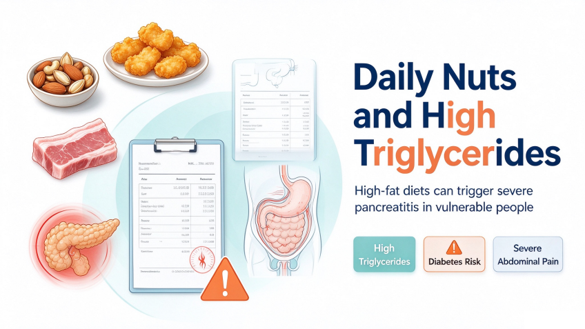
Man lands in ICU with sky-high blood lipid levels after daily nut consumption

Man lands in ICU with sky-high blood lipid levels after daily nut consumption

Mr. Fang (a pseudonym), 41, never imagined that simply eating an extra handful of nuts would land him in the intensive care unit (ICU) and necessitate emergency interventions, including hemofiltration. The culprit behind this crisis was acute pancreatitis triggered by hyperlipidemia—a condition brought on by a high-fat intake—which, compounded by his existing underlying health issues, made his situation exceptionally perilous.

On the day of the incident, shortly after consuming a large quantity of nuts, Mr. Fang began experiencing persistent upper abdominal pain. His family rushed him to Xi’an Gaoxin Hospital, where CT scans and amylase level tests confirmed a diagnosis of acute pancreatitis. Further examination revealed that his triglyceride levels had skyrocketed to 82.40 mmol/L (the normal range is typically <1.7 mmol/L). He was also suffering from diabetic ketoacidosis and hyperglycemia. Given his medical history—which includes coronary heart disease (with prior coronary stent placement) and type 2 diabetes—his case presented a complex scenario of multiple pre-existing systemic conditions combined with an acute, severe illness.

Physicians in the Department of Gastroenterology at Xi’an Gaoxin Hospital identified the condition as “hyperlipidemia-induced pancreatitis”—a type often triggered by overeating or the excessive consumption of high-fat foods. With his triglyceride levels dangerously high, immediate lipid-lowering intervention was essential to prevent the pancreatic inflammation from worsening and becoming life-threatening. Because medication alone would not act quickly enough, Mr. Fang was transferred to the ICU for hemofiltration—a critical procedure used to rapidly clear triglycerides from the bloodstream. After three days of intensive care, his triglyceride levels plummeted from an off-the-charts 82.40 mmol/L to 2.99 mmol/L. His ketoacidosis was resolved, and his abdominal pain subsided, allowing him to be moved back to a general ward.

Just as it seemed he was on the mend, however, Mr. Fang attempted to take a sip of water—only for the abdominal pain to flare up once again, accompanied by a high fever. A follow-up CT scan revealed that Mr. Fang’s pancreatitis had progressed to Balthazar grade D. There was increased peripancreatic exudation, the inflammation had spread to the adjacent intestines, and a secondary infection had developed. The gastroenterology team promptly adjusted his treatment plan. With careful management, Mr. Fang’s abdominal pain was brought under control; he was eventually able to resume eating without any recurrence of pain and was successfully discharged.

Following his discharge, Mr. Fang has strictly adhered to a low-fat diet and consistently taken his lipid-lowering medication. Subsequent outpatient check-ups have shown his triglyceride levels remaining within the normal range, and his condition has stayed stable.

The Department of Gastroenterology at Xi’an Gaoxin Hospital notes that autumn and winter are peak seasons for acute pancreatitis. Individuals with underlying health conditions, in particular, should be wary of the risks posed by overindulging in food. Please keep the following four points in mind:

High-fat diets are “silent killers”:

While high-fat foods—such as nuts, fatty meats, and fried items—may be delicious, consuming them in excess can trigger hyperlipidemia-induced pancreatitis. Those who already have high blood lipid levels should exercise particular restraint.

Higher risk for those with underlying conditions:

Individuals with pre-existing conditions such as diabetes, coronary heart disease, or hyperlipidemia already have more vulnerable pancreatic function and metabolic systems. They should avoid overeating, stick to a light, low-fat diet, and regularly monitor relevant health indicators.

Do not “tough out” abdominal pain:

Sudden, persistent upper abdominal pain—accompanied by symptoms like bloating or nausea—could be a sign of acute pancreatitis. Seek medical attention immediately; delaying treatment can lead to a rapid deterioration of the condition.

Dietary care during recovery:

During recovery from pancreatitis, patients should follow a gradual dietary progression: starting with water, then moving to clear liquids, semi-liquids, and finally soft foods. Do not resume eating or drinking too soon, as this could irritate the pancreas and trigger a recurrence of inflammation.

Although hyperlipidemia-induced acute pancreatitis is a serious and dangerous condition, it can be effectively managed through timely medical intervention, evidence-based treatment, and long-term health management following discharge. Do not let a fleeting craving for food cause irreversible harm to your body. This is especially critical for those with underlying medical conditions, who must take to heart the importance of “controlling one’s diet and following medical advice.”

Digestive Disease Hospital, Xi’an Gaoxin Hospital

The Digestive Disease Hospital at Xi’an Gaoxin Hospital is a specialized team dedicated to the clinical study and treatment of digestive system disorders. The department provides long-term diagnostic and therapeutic services for both common and complex, severe digestive diseases in Xi’an and its surrounding regions. Through years of medical practice, the team has developed a unique clinical approach and accumulated extensive experience in managing a wide range of digestive conditions—from common ailments to complex, life-threatening cases—earning high praise from patients and their families.

Led by President Song Ying, who serves as the head of the Digestive Disease Hospital and the discipline leader, the department comprises two inpatient wards, a day-surgery unit, an endoscopy center, a laboratory, and a gastrointestinal rehabilitation center. There are 160 inpatient beds available. The endoscopy center spans 1,500 square meters and features 10 standard procedure rooms equipped with state-of-the-art technology, supported by a well-structured team of medical professionals. Key specialized procedures offered by the department include endoscopic treatment for acute gastrointestinal bleeding, endoscopic management of esophageal and gastric varices, percutaneous endoscopic gastrostomy (PEG), endoscopic sclerotherapy and band ligation for internal hemorrhoids, and endoscopic resection of gastrointestinal tumors. The Department of Gastroenterology is a key medical discipline in Xi’an and a flagship department within the hospital, boasting robust clinical expertise. It currently serves as the chair institution for four academic organizations: the Digestive Disease Prevention and Treatment Professional Committee of the Shaanxi Healthcare Society, the Gastroenterology and Digestive Endoscopy Professional Committee of the Shaanxi Non-Public Medical Institutions Association, the Digestive Endoscopy Professional Committee of the Shaanxi Research-Oriented Hospital Society, and the Digestive Endoscopy Professional Committee of the Shaanxi Association of Integrated Traditional Chinese and Western Medicine. Furthermore, it acts as a vice-chair unit for both the Esophageal and Gastric Varices Study Group and the Internal Hemorrhoids Collaborative Group under the Chinese Society of Digestive Endoscopy (CSDE). The department is also designated as a training and demonstration base for digestive endoscopy by the Chinese Non-Public Medical Institutions Association, a training center for the sclerotherapy of esophageal and gastric varices under the Chinese Medical Association, a standardized training base for the prevention and treatment of gastrointestinal bleeding in Shaanxi Province, and a governing member institution of the Asia-Pacific Portal Hypertension Alliance.

Specialized Diagnostic and Therapeutic Techniques

Carbon-13 (13C) urea breath test, food intolerance testing, transient elastography (FibroScan) for liver fibrosis, psychological assessment, routine and painless (sedated) gastrointestinal endoscopy, endoscopic chromoendoscopy for early-stage cancer detection, advanced diagnostic imaging (NBI, BLI, and LCI), endoscopic ultrasound (EUS), emergency endoscopic hemostasis, removal of foreign bodies from the digestive tract, endoscopic variceal ligation (EVL) and sclerotherapy for esophageal varices, tissue adhesive injection (glue embolization) for gastric variceal bleeding, endoscopic mucosal resection (EMR) and argon plasma coagulation (APC) for polyps, stent placement for benign or malignant esophageal and biliary strictures, endoscopic retrograde cholangiopancreatography (ERCP), endoscopic sphincterotomy (EST) for stone extraction, endoscopic nasobiliary drainage (ENBD), biliary stent placement, percutaneous endoscopic gastrostomy (PEG), endoscopic placement of enteral nutrition tubes, placement of intestinal obstruction decompression tubes, endoscopic submucosal dissection (ESD), endoscopic submucosal excavation (ESE), endoscopic full-thickness resection (EFTR), submucosal tunneling endoscopic resection (STER), and peroral endoscopic myotomy (POEM).

globalmedpool.com

An Overview of Small, Benign Liver Lesions

An Overview of Small, Benign Liver Lesions

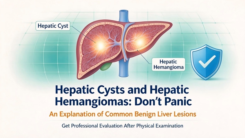
In routine medical check-ups, people are sometimes surprised to find reports of abnormal liver lesions, with “hepatic cysts” and “hepatic hemangiomas” being among the most common findings. These terms can be alarming, often leading to concerns about potential malignancy. The Radiology Department at Xi’an Gaoxin Hospital provides an overview of these two benign liver conditions to help the public better understand these “minor issues.”

 Hepatic Cysts: “Small Fluid-Filled Sacs” in the Liver

A hepatic cyst is a very common benign liver condition. In radiological imaging, it appears as a well-defined, homogeneous, low-density lesion—essentially a small, fluid-filled sac within the liver tissue. The sac contains clear, sterile serous fluid and has no connection to the intrahepatic bile ducts; therefore, it does not interfere with the normal secretion of bile.

Hepatic cysts are categorized as either congenital or acquired. Congenital cysts are generally believed to originate from developmental abnormalities of the intrahepatic bile ducts during the embryonic stage, while acquired cysts are often triggered by factors such as trauma, inflammation, or parasitic infections.

The vast majority of hepatic cysts cause no noticeable discomfort and are typically discovered incidentally during routine check-ups. Small, asymptomatic cysts generally require no specific treatment—periodic follow-up and observation are usually sufficient. If a cyst exceeds 5 centimeters in diameter and begins to exert pressure on surrounding organs, it may cause mild symptoms such as abdominal bloating, a dull ache in the upper right abdomen, or a loss of appetite. When it is necessary to distinguish a hepatic cyst from other liver lesions, CT or MRI scans can provide a clear diagnostic basis. A hallmark of a typical hepatic cyst on imaging is its smooth, well-defined border and uniform density; notably, it does not show “enhancement” (increased brightness) following the administration of a contrast agent—a key feature for identification.

Hepatic Hemangiomas: “Clusters of Blood Vessels” in the Liver

A hepatic hemangioma is the most common benign liver tumor, with cavernous hemangiomas accounting for the vast majority of cases. On diagnostic imaging, these lesions appear as well-defined masses within the liver, composed of a dense network of dilated, tiny blood vessels—resembling a tangled web of vessels. They are soft in texture, grow slowly, and do not invade surrounding healthy tissue.

The exact pathogenesis of hepatic hemangiomas remains not fully understood; however, the prevailing view is that they are linked to congenital vascular developmental abnormalities. Clinical research indicates that hepatic hemangiomas may tend to enlarge during periods of significant hormonal fluctuation—such as puberty or pregnancy—suggesting that hormonal changes may influence the lesions.

Most individuals with hepatic hemangiomas are asymptomatic. For lesions smaller than 5 cm that cause no discomfort, routine follow-up and observation are generally sufficient. If a lesion exceeds 5 cm in diameter, causes symptoms due to compression of adjacent structures, or is located beneath the liver capsule—posing a risk of rupture—it is necessary to seek medical attention so that a physician can determine an appropriate management plan. CT or MRI scans typically reveal characteristic enhancement patterns, which help clinicians distinguish hepatic hemangiomas from malignant liver tumors.

 How to Manage a Diagnosis of Hepatic Cysts or Hepatic Hemangiomas

First and foremost, avoid excessive anxiety. Both hepatic cysts and hepatic hemangiomas are benign lesions characterized by slow growth. There is no reliable evidence to suggest that hepatic hemangiomas carry a risk of malignant transformation, and congenital hepatic cysts also grow very gradually; the vast majority of patients remain entirely asymptomatic throughout their lives.

If such lesions are detected during a medical check-up, the core principle is to follow the recommendations provided by radiologists and relevant specialists. For small lesions, the standard approach is periodic monitoring to track any changes. If a lesion meets the criteria for clinical intervention, a physician will evaluate the situation and determine the most appropriate course of action—there is no need for indiscriminate surgery or medication.

Arthroscopic subacromial balloon implantation

Arthroscopic subacromial balloon implantation

Arthroscopic subacromial balloon implantation

Even buttoning a shirt had become an impossible task—for the past few years, 67-year-old Mr. L had been plagued by persistent shoulder pain. Although he had previously undergone arthroscopic rotator cuff repair on his right shoulder, a subsequent injury left him with restricted movement in that shoulder for the past six months.

An MRI scan revealed a massive rotator cuff tear (defined as a tear larger than 5 cm, or the complete rupture of two or more rotator cuff tendons), accompanied by significant muscle degeneration.

The sports medicine team at Wanying Orthopedics diagnosed Mr. L’s condition as an “irreparable massive rotator cuff tear,” meaning the tissue could no longer be repaired. Following an in-depth multidisciplinary discussion—and noting that the patient had not yet developed osteoarthritis—the team opted for an advanced procedure: the implantation of an InSpace subacromial balloon.

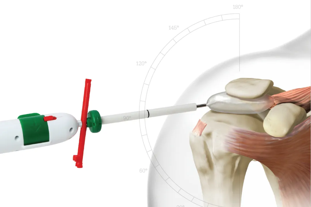
Supported by a skilled anesthesia team, the surgeons completed the balloon implantation in just 30 minutes. By placing a small, fluid-filled balloon between the acromion and the humeral head, they effectively restored the “fulcrum” for shoulder movement, re-establishing the balance of the joint’s force couples. Furthermore, the balloon prevents friction between the acromion and the humeral head, helping to halt further joint degeneration.

The surgery was a success, and Mr. L experienced immediate relief from his shoulder pain. “During the morning rounds, I was actually able to lift my arm to say hello!” he exclaimed. “Seeing such an ‘instant’ result has completely convinced me of the clinical expertise at Wanying Medical.” Mr. L was discharged the following day and will soon begin his post-operative rehabilitation.

Overcoming the Challenge of Age-Related Shoulder Pain
Yaoying Continues to Introduce Innovative Minimally Invasive Treatment Techniques

As the global population ages, rotator cuff injury has become a common clinical shoulder disorder. The condition most frequently stems from the gradual wear and tear of tendon tissue over time; without timely intervention, it often progresses to a massive rotator cuff tear. Such tears result in shoulder weakness and pain, hindering the performance of many daily activities—such as combing one’s hair or getting dressed—and causing nighttime pain severe enough to disrupt sleep.

Pain Points of Traditional Treatments

⚠️ Significant Trauma: Open surgery requires cutting the deltoid muscle, necessitating a long period of postoperative immobilization.
⚠️ High Risk: The rate of re-tearing following surgery for massive tears exceeds 40%.

Advantages of InSpace Balloon Technology

✔️ Innovative “Water Balloon” Technology: A biodegradable balloon is implanted to act as a cushion, replacing the function of a torn tendon and reducing friction.
✔️ Millimeter-Level Precision: Precise, arthroscopic placement avoids damage to nerves and blood vessels, with no disruption to the bony structures of the shoulder joint.
✔️ Rapid Recovery: Patients are typically discharged the day after surgery, with a recovery period 50% shorter than that of traditional surgery.
✔️ Minimally Invasive: The procedure is performed through three 5mm incisions, with blood loss of less than 10ml.
✔️ High Safety Profile: Since receiving its CE mark in 2010, the InSpace bioabsorbable shoulder balloon has been used to successfully treat over 28,000 patients across North America, the European Union, and other regions.
✔️ Reversible: The balloon degrades naturally over two years, leaving the option open for future surgical interventions.

Who is a candidate for the InSpace subacromial balloon implant procedure?

  • Patients with massive, irreparable rotator cuff tears—specifically those with large tears (>5 cm) that cannot be repaired through conventional surgical techniques.
  • Patients with mild to moderate glenohumeral osteoarthritis; the balloon implant can help alleviate pain caused by arthritis and improve shoulder function.
  • Patients aged 65 and older; compared to traditional surgery, the InSpace balloon implant procedure can offer faster functional recovery and pain relief.

Please note: Determining whether the InSpace balloon implant procedure is appropriate for you requires an in-person consultation at a hospital. A physician will conduct a comprehensive assessment based on your specific medical condition, overall physical health, and other relevant factors, and will then work with you to discuss and determine the most suitable treatment plan.

Proton Therapy: An Excellent Treatment Option for Giant Hepatic Hemangiomas

Proton Therapy: An Excellent Treatment Option for Giant Hepatic Hemangiomas

Proton Therapy: An Excellent Treatment Option for Giant Hepatic Hemangiomas

Most hepatic hemangiomas grow slowly and typically do not cause obvious clinical symptoms; they are usually detected during routine health check-ups or abdominal ultrasound and CT scans performed for other conditions.

In some patients, the hemangioma is large enough to compress adjacent organs—such as the gastrointestinal tract—leading to symptoms like upper abdominal discomfort, distension and pain, indigestion, nausea, and vomiting. In very rare instances, the hemangioma may rupture, triggering critical abdominal complications such as intraperitoneal hemorrhage.

Upon detection, the preferred clinical approach is regular follow-up to monitor changes in the tumor’s size and morphology, with timely intervention initiated should any abnormalities arise.

Treatment becomes necessary when the hemangioma exceeds 5 cm in diameter or causes clinical symptoms; when it continues to enlarge, compressing surrounding organs or adversely affecting the cardiovascular system; or when there is a risk of rupture, hemorrhage, or life-threatening complications. Current mainstream treatment modalities include surgical resection, interventional therapy, and radiotherapy.

 46-Year-Old Woman Diagnosed with Giant Hepatic Hemangioma; Blood Transfusion Contraindication Complicates Surgery

Literature reports a case involving the use of proton therapy for a giant hepatic hemangioma. Researchers note that proton therapy is safe for treating hepatic hemangiomas, achieves ideal local control, causes no severe liver damage, and minimizes the risk of long-term adverse effects.

The patient was a 46-year-old woman with normal liver function. Elevated hepatobiliary enzyme levels were noted during a routine check-up, and an abdominal ultrasound revealed a hepatic mass. An abdominal CT scan showed an 80 × 80 mm lesion in the left lobe of the liver; combined with contrast-enhanced imaging, the lesion was confirmed to be a giant hepatic hemangioma.

Imaging also revealed peripheral bile duct dilation, with the bile ducts and blood vessels compressed by the tumor. Physical examination showed a flat, soft abdomen with tenderness in the right costal region. Comprehensive assessment indicated that the giant hepatic hemangioma required prompt intervention.

 Continued Tumor Shrinkage and Sustained Remission for 15 Years After Proton Therapy

Due to religious restrictions preventing blood transfusions—which made surgical resection difficult to perform—the patient was referred for proton therapy. Initial assessment indicated Child-Pugh Class A liver function (suggesting good function or only mild impairment) and no elevation in tumor markers.

The patient underwent proton therapy with a total dose of 28.6 Gy delivered in 13 fractions. Following treatment, the tumor continued to shrink: it measured 50×50 mm after one year, 44×33 mm at 1.5 years, 40×26 mm at two years, and 36×26 mm at three years. Annual ultrasound follow-ups showed continued tumor regression.

An MRI scan 11 years post-treatment revealed the lesion had shrunk to 29×15 mm; the lesion appeared stable and was difficult to identify via ultrasound. Aside from localized atrophy in the irradiated area, the remaining liver structure remained intact, with no hepatic deformity. Follow-up blood tests showed no elevation in hepatobiliary enzymes, and symptoms of abdominal discomfort had completely resolved. The patient achieved a performance status score of 0, maintained a normal lifestyle, and attained clinical recovery. Ultrasound and imaging follow-ups confirmed that the state of remission has been sustained for 15 years.

Complete resolution of liver metastases from esophageal cancer following 30 sessions of proton therapy.

Complete resolution of liver metastases from esophageal cancer following 30 sessions of proton therapy.

Complete resolution of liver metastases from esophageal cancer following 30 sessions of proton therapy.

Proton Therapy for Liver Cancer: 5-Year Local Progression-Free Rate Exceeds 85%

Among men under the age of 60, liver cancer ranks first in terms of cancer incidence and mortality.

Deaths among cancer patients are largely caused by local recurrence and distant metastasis. The liver is the body’s largest solid organ and has a rich blood supply; consequently, primary liver tumors are prone to intrahepatic or extrahepatic metastasis, and the liver is also a common site for metastasis from many other malignancies.

Metastatic liver cancer—also known as secondary liver cancer—refers to tumors formed when malignancies from other parts of the body spread to the liver. During the course of malignant disease, 25% to 50% of primary tumors metastasize to the liver. Statistically, liver metastasis is the second most common form of spread, ranking just behind lymphatic metastasis. Metastases originating from the digestive tract and pelvic region are particularly common, involving sites such as the stomach, small intestine, colon, rectum, gallbladder, pancreas, prostate, uterus, and ovaries; however, metastases from the thyroid, breast, nasopharynx, lungs, and kidneys are also frequently observed.

Proton Therapy for Liver Cancer: 5-Year Local Progression-Free Rate Exceeds 85%

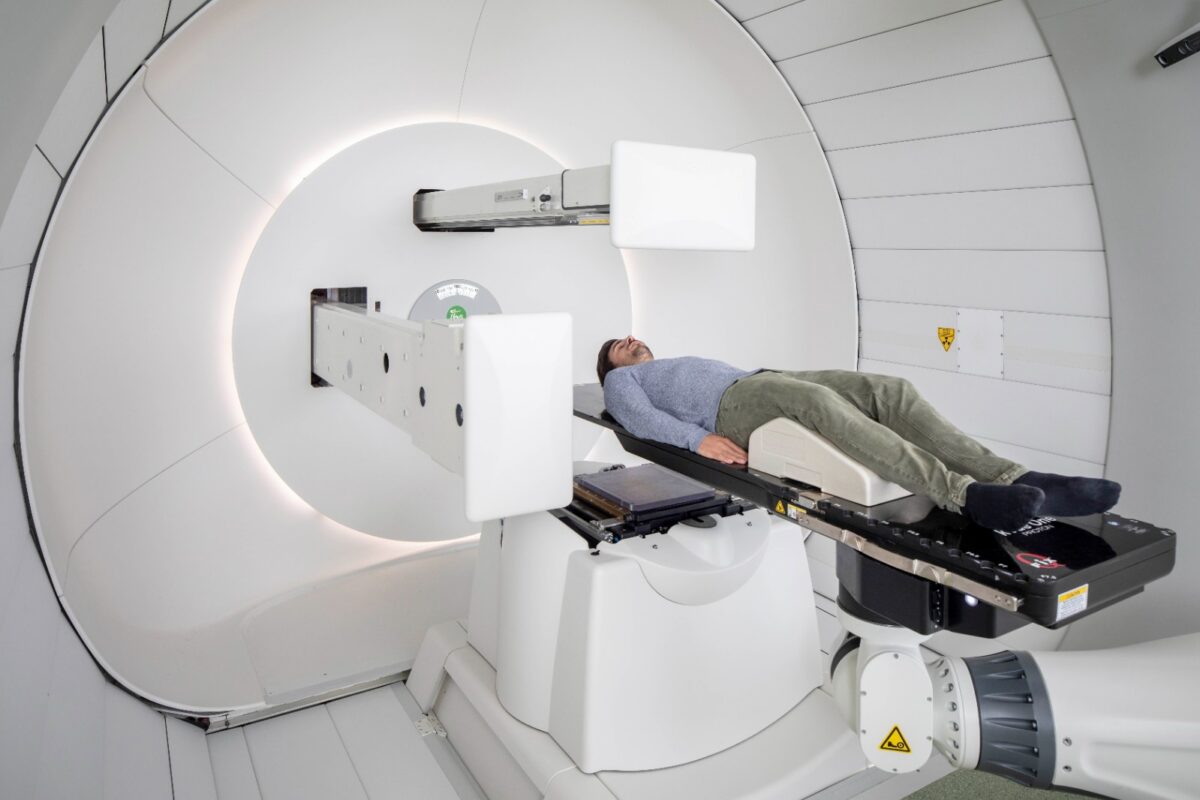
Surgery is the preferred treatment for most early-stage liver cancers, yet the majority of patients are ineligible for surgery at the time of diagnosis. Radiation therapy serves as a crucial treatment option for unresectable liver cancer. Liver cancer cells are relatively sensitive to radiation; however, the low radiation tolerance of normal liver tissue limits the use of X-ray radiotherapy, particularly in patients with multiple liver tumors. Proton therapy allows for the delivery of high radiation doses to the tumor while significantly reducing the dose received by surrounding normal tissues.

Compared to X-ray therapy, proton therapy offers superior protection for normal liver tissue. Studies indicate that proton therapy yields favorable outcomes for primary hepatocellular carcinoma, achieving a 5-year local progression-free rate of over 85%. Additionally, research suggests that proton therapy is associated with a reduction in adverse events. In a Phase II trial, acute toxicity during proton therapy was minimal, consisting only of Grade 1 erythema and mild fatigue; no acute toxicities interfered with the proton therapy.

A 53-year-old male diagnosed with esophageal cancer and three liver metastases

A case of proton therapy for liver metastases from esophageal cancer was previously reported in the journal International Surgery.

A 53-year-old male presented with complaints of heartburn and a choking sensation. Esophagogastroduodenoscopy revealed cancer at the esophagogastric junction and in the upper thoracic esophagus. Additionally, computed tomography (CT) scans showed multiple liver metastases and mediastinal lymph node metastasis. Specifically, there were three liver metastases (measuring 45.5 mm in segment S2, 67.5 mm in S4, and 48.3 mm in S6) and one mediastinal lymph node metastasis. Routine laboratory test results, including tumor marker levels, were normal. Positron emission tomography (PET) showed fluorodeoxyglucose (FDG) accumulation in the liver (segments S2, S4, and S6), the primary tumor sites (esophagogastric junction and upper thoracic esophagus), and a left supraclavicular lymph node. Based on these findings, the patient was diagnosed with Stage IV disease (T3N1M1 [liver] for the lower thoracic esophageal cancer and T3 for the upper thoracic esophageal cancer) and a chemotherapy regimen was planned.

Over 30 proton therapy sessions: Complete elimination of all tumors

The efficacy of chemotherapy was assessed using the World Health Organization (WHO) criteria for response in solid tumors. However, disease progression was observed after four cycles of chemotherapy. Consequently, the treatment plan was modified to combine proton therapy with chemotherapy. Proton beams were delivered via two oblique ports. Initially, the primary tumors, the left supraclavicular lymph node, and the liver metastasis in segment S6 were treated with proton therapy—receiving 31 fractions (total dose: 68.2 Gy) and 30 fractions (total dose: 66 Gy), respectively. Subsequently, chemotherapy was administered. Finally, the liver metastases in segments S2 and S4 were treated with proton therapy, receiving 30 fractions for a total dose of 66 Gy. Approximately two months after the completion of proton therapy combined with chemotherapy, PET scans showed the disappearance of FDG uptake in all liver metastasis sites, the primary tumor, and the mediastinal lymph nodes.

CT and PET imaging confirmed the complete resolution of all target lesions—specifically those in liver segments S2, S4, and S6, as well as the upper mediastinal lymph nodes. Esophagogastroduodenoscopy revealed the resolution of non-target lesions (the primary tumor located at the upper thoracic esophagus and the gastroesophageal junction), indicating a complete remission of the esophageal cancer. No treatment-related acute or late toxicities were observed.

In summary, proton therapy is a highly advanced medical technology that yields excellent results in treating both primary and metastatic liver cancer, offering new hope to cancer patients today.

Raffles Hospital

Opening a lifeline for acute stroke patients

In the race for life, time is of the essence; every second can determine the difference between life and death. Recently, the team led by Professor Chi Luxiang at the Chongqing Raffles Pan-Vascular Disease Management Center executed a dramatic life-saving intervention, successfully performing surgery on Mr. Xie, a 66-year-old patient suffering from an acute stroke, and pulling him back from the brink of death.

Critical Moment: Stroke Strikes, Life Hanging by a Thread

Mr. Xie suddenly experienced limb weakness and slurred speech and was rushed to Chongqing Raffles Hospital. Emergency imaging assessments confirmed a diagnosis of acute ischemic stroke. By this time, the patient had already missed the optimal time window for intravenous thrombolysis, rendering standard thrombolytic therapy unfeasible. With the blood vessel remaining occluded and brain tissue suffering progressive ischemic necrosis, the patient faced an imminent risk of massive cerebral infarction and cerebral herniation—conditions that could prove fatal.

Acute stroke treatment relies on a strict “golden window” for intervention. Once a large vessel is occluded, conservative treatment after this window passes yields a very poor prognosis, with significantly higher rates of disability and mortality. Faced with this complex clinical challenge, Professor Chi Luxiang’s team immediately activated the stroke emergency “green channel.” They rapidly conducted a multidisciplinary consultation to comprehensively assess vascular conditions and the extent of cerebral ischemia, subsequently formulating an interventional surgical plan.

Precision Intervention: Minimally Invasive Surgery Saves the Day

The team decided to perform **thrombus aspiration and carotid artery stenting** for Mr. Xie. This type of minimally invasive neuro-interventional procedure requires no craniotomy—leaving only a tiny puncture wound—but demands exceptional precision and diagnostic skill from the surgical team.

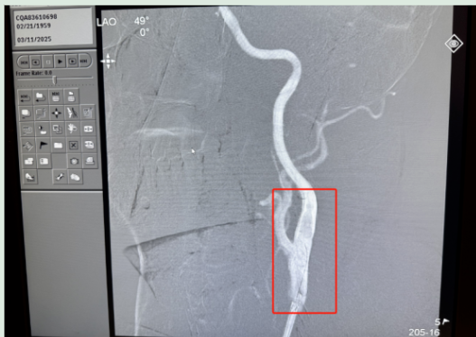
During the procedure, guided by digital subtraction angiography (DSA) equipment, Professor Chi and his team navigated a micro-catheter from the femoral artery into the intracranial space. They precisely reached the site of the occlusion, successfully aspirated and removed the obstructing thrombus, and simultaneously implanted a stent into the narrowed carotid artery to restore blood flow to the brain. The surgery was completed successfully; the occluded vessel was reopened, and blood supply to the brain tissue was rapidly restored.

Following the procedure, the patient was transferred to the Intensive Care Unit (ICU) for close monitoring. Following collaborative management by the critical care, neurology, neurosurgery, and nursing teams, the patient’s limb strength and speech function gradually recovered; the patient was successfully transferred out of the intensive care unit and, after undergoing subsequent rehabilitation, returned to a normal life.

Proton Therapy Precisely Targets "Double Primary Cancers": Prostate Lesion Inactive; Rectosigmoid Junction Cancer Locally Eradicated

Proton Therapy Precisely Targets “Double Primary Cancers”: Prostate Lesion Inactive; Rectosigmoid Junction Cancer Locally Eradicated

Proton Therapy Precisely Targets “Double Primary Cancers”: Prostate Lesion Inactive; Rectosigmoid Junction Cancer Locally Eradicated

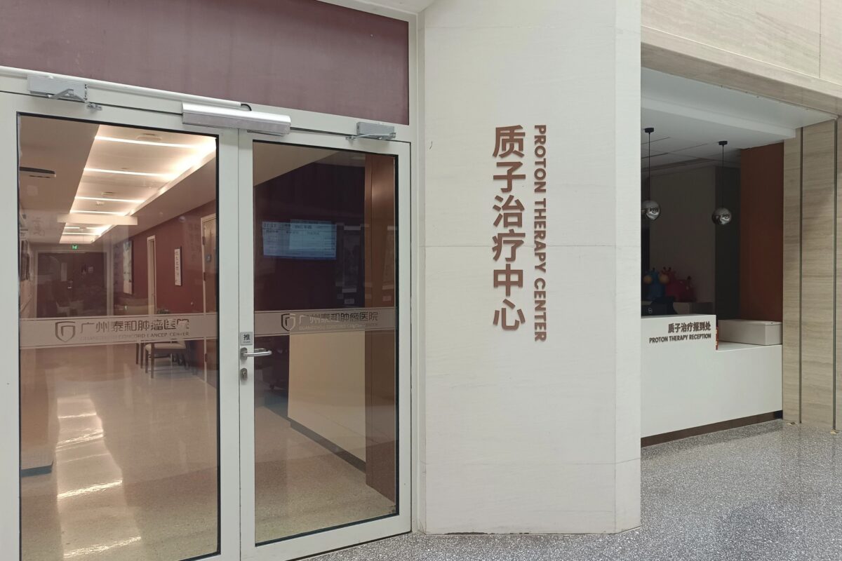
Mr. C, now in his sixties—an age when he should have been enjoying his retirement—was diagnosed with both prostate cancer and cancer at the rectosigmoid junction. Faced with this sudden storm of adversity, Mr. C did not hesitate to seek active medical treatment. After consulting various sources, he came to Guangzhou Concord Cancer Center for proton therapy.

Fortunately, the personalized treatment plan tailored by the hospital’s multidisciplinary team yielded inspiring results: the prostate cancer lesion shrank, tumor markers returned to normal levels, and imaging showed no signs of residual activity. Furthermore, surgery confirmed a pathological complete response for the rectosigmoid junction cancer, marking a decisive victory in this battle against dual malignancies.

Diagnosed with double primary cancers: seeking the “globally optimal solution”

During a routine health check-up, Mr. C received an alarming result: his total prostate-specific antigen (TPSA) level had skyrocketed to more than ten times the upper limit of normal. Further examinations confirmed a diagnosis of high-risk, locally advanced prostate cancer that had already invaded the seminal vesicles.

For cases of this nature, surgery often fails to completely eradicate the tumor, and the risk of postoperative recurrence is high. The National Comprehensive Cancer Network (NCCN) guidelines explicitly recommend “external beam radiotherapy combined with long-term androgen deprivation therapy” as a top-tier treatment option for prostate cancer classified as T3b or higher (indicating tumor invasion into the seminal vesicles or beyond).

Given the prostate’s unique anatomical location—in close proximity to the bladder, small intestine, and rectum—traditional photon radiotherapy frequently affects these normal tissues. This can lead to adverse effects such as radiation enteritis, diarrhea, rectal bleeding, or hematuria, severely compromising the patient’s quality of life. With advancements in medical technology, proton therapy has emerged as a highly effective treatment for prostate cancer. Studies indicate that the “Bragg peak” effect of proton therapy can reduce radiation exposure to organs near the prostate by 80%, thereby minimizing radiation to the bladder, intestines, and surrounding healthy tissue; furthermore, men undergoing proton therapy face a significantly lower risk of side effects such as sexual dysfunction and urinary incontinence.

Seeking proton therapy, Mr. C traveled to Guangzhou Taihe Cancer Hospital. Unexpectedly, a comprehensive follow-up examination revealed a second primary cancer: a rectosigmoid junction carcinoma.

Faced with the complex scenario of dual primary cancers—prostate cancer and rectosigmoid junction carcinoma—both located in the pelvic region, the proton therapy team at Guangzhou Taihe Cancer Hospital convened a multidisciplinary discussion involving experts from surgery, medical oncology, radiology, and the endoscopy center. Moving beyond the traditional “one cancer, one treatment” approach, they adopted a strategy of treating distinct diseases simultaneously, proposing an integrated concurrent chemoradiotherapy plan. Taking into account the patient’s multiple underlying medical conditions and personal treatment preferences, the medical team devised a comprehensive treatment plan combining proton therapy and concurrent chemotherapy to precisely target both primary cancer sites. The specific strategy involved administering curative-intent proton radiotherapy combined with endocrine therapy for the prostate cancer. Simultaneously, the radiation field encompassed the tumor at the rectosigmoid junction and its associated lymphatic drainage areas, accompanied by concurrent chemotherapy; the aim was to shrink the rectosigmoid tumor and downstage the disease, thereby creating favorable conditions for subsequent surgery and increasing the likelihood of a successful curative resection.

“In complex cases like this, the key lies in emphasizing an integrated strategy rather than simply layering treatments,” explained Dr. Luo Yingshan from the Department of Radiation Oncology. “We need to balance the timing of treatments for both cancers against the risk of side effects. Precision technologies like proton therapy allow us to effectively cover multiple target areas while better protecting shared organs at risk surrounding the lesions, providing crucial support for achieving the dual goals of curative treatment and the preservation of quality of life.”

Proton therapy creates a surgical window; postoperative pathology confirms ideal therapeutic efficacy.

As the first proton therapy center in South China to enter clinical operation, the Proton Therapy Center at Guangzhou Taihe Cancer Hospital strictly adheres to the radiotherapy guidelines and standard protocols of MD Anderson Cancer Center, dedicated to providing patients with personalized, cutting-edge treatment plans. The center is equipped with the world-leading Varian ProBeam® proton therapy system and led by authoritative domestic experts; the team possesses extensive experience, having managed thousands of clinical cases in the field of proton therapy.

After completing 28 sessions of proton therapy, Mr. C continued his chemotherapy at Guangzhou Taihe Cancer Hospital. Follow-up imaging three months later showed no signs of activity in his prostate lesion. Meanwhile, endoscopic examination revealed no obvious mass at the rectosigmoid junction, though surgery was still required for definitive confirmation.

Following a comprehensive re-evaluation by the multidisciplinary team, it was determined that Mr. C met the criteria for surgery. A surgical team led by Associate Chief Physician Xie Xiaoming performed a “laparoscopic-assisted radical resection of rectosigmoid junction cancer.” The procedure went smoothly with intraoperative blood loss of less than 10 ml, and the patient recovered well postoperatively. Even more encouragingly, postoperative pathology confirmed that the patient had achieved a pathological complete response (pCR).

Associate Chief Physician Xie Xiaoming further explained: “pCR is tantamount to a clinical cure and represents the ideal goal of neoadjuvant or conversion therapy. It indicates a very low risk of tumor recurrence and a favorable long-term prognosis. This also suggests that this comprehensive treatment model—centered on proton therapy—holds broad clinical prospects for similar cases.”

Today, Mr. C is recovering well and gradually returning to his normal life. This case powerfully demonstrates that when facing the complex scenario of double primary cancers—prostate cancer and rectosigmoid junction cancer—located in close proximity within the pelvic cavity, a personalized, integrated treatment plan formulated by a multidisciplinary team, combined with advanced technologies like proton therapy, can effectively control or cure both cancers, thereby delivering genuine survival benefits and ensuring a good quality of life for the patient.

globalmedpool

The Five-Year CAR-T Marathon of an 11-Year-Old Girl with Acute B-Cell Lymphoblastic Leukemia

Five years after sequential CAR-T therapy, the pediatric patient has achieved clinical cure—a ray of hope after nine years of waiting.

After undergoing sequential CAR-T therapy, the young patient Xiaoxiao achieved clinical remission and was officially declared free of leukemia—the outcome she and her family had awaited for nine years.

In the immediate aftermath of the CD19 CAR-T cell infusion, no one could predict whether this frail, slight girl would successfully weather the ordeal and make a full recovery.

Diagnosis, chemotherapy, discontinuation of treatment, and subsequent disease recurrence.

Xiao Xiao’s complexion deteriorated and she began crying incessantly for no apparent reason, prompting a visit to a local hospital. Bone marrow cytology and immunophenotyping confirmed a diagnosis of acute B-lymphoblastic leukemia (B-ALL); she was just over two years old at the time.

For the next three-plus years, chemotherapy, bone marrow aspirations, and intrathecal injections dominated much of her childhood. Fortunately, repeated bone marrow assessments consistently showed negative results. She completed her chemotherapy regimen, and the family believed the hardest part was over. However, a follow-up bone marrow examination shattered that peace: 25.6% of the cells in her bone marrow were identified as primitive, immature lymphocytes, indicating a full-blown relapse of the leukemia.

To this day, Xiao Xiao’s father vividly remembers the moment he received that follow-up report. The sudden news made him feel, for a time, as though the perseverance of the past few years had been in vain—yet he had no time to give in to despair. He gathered all her medical records and began actively seeking avenues for continued treatment.

Traveling to Beijing amidst the pandemic to seek treatment from Director Pan Jing’s team.

The journey to seek medical treatment across provinces was fraught with obstacles. During a relapse, every day of delay risked a continued increase in tumor burden. Without hesitation, Xiaoxiao’s father took the necessary protective measures and rushed with her from their hometown to Beijing.

At the Department of Hematology-Oncology and Immunotherapy at Beijing Gaobo Hospital, Director Pan Jing’s team meticulously reviewed Xiaoxiao’s entire medical history—spanning her initial diagnosis, the period off medication, and the subsequent relapse—and conducted a comprehensive assessment based on her current physical condition. After thorough consultation with the family, the team determined that Xiaoxiao met the eligibility criteria for CAR-T cell therapy.

“The child’s baseline physical condition is reasonably good; following chemotherapy after the relapse, her tumor burden has decreased, making her a suitable candidate for sequential therapy.” Director Pan’s assessment brought the first sense of relief to Xiaoxiao’s father, easing the anxiety that had been weighing heavily on him. The medical team subsequently devised a treatment plan combining tumor-burden reduction with sequential CD19/CD22 CAR-T cell therapy.

CAR-T is not a single procedure, but a complete, continuous treatment cycle.

The public often mistakenly believes that CAR-T cell therapy consists solely of the cell infusion itself; however, the infusion is merely one component of the entire treatment process.

The comprehensive process begins with a preliminary assessment, during which doctors evaluate the patient’s physical condition, disease progression, and treatment history to determine suitability for CAR-T therapy. This is followed by the formulation and dynamic adjustment of a personalized treatment plan, encompassing key steps such as cell manufacturing and infusion. Post-infusion, patients must manage various risks—including fever, inflammatory responses, infections, and immune reconstitution—while undergoing long-term follow-up to continuously monitor therapeutic efficacy and physical recovery. These stages are interconnected and interdependent; they cannot be viewed in isolation.

For the patient, CAR-T represents a continuous, comprehensive treatment journey rather than a single surgical procedure. The ultimate outcome is rarely determined by the day of infusion alone but is instead shaped by the management of the entire clinical process.

Xiaoxiao first underwent chemotherapy to reduce tumor burden and received FC pre-conditioning, successfully completing the CD19 CAR-T cell infusion. Follow-up assessments showed no abnormal lymphocytes regarding minimal residual disease (MRD) in the bone marrow, and morphological results for both bone marrow and peripheral blood were negative. Subsequently, in accordance with the established plan, she completed the sequential infusion of autologous human-origin CD22 CAR-T cells.

Following the cell infusions, Xiaoxiao experienced fever and a period of bone marrow suppression, and she also contracted COVID-19 during the later stages of the sequential treatment. Amidst the harsh winter of the country’s first major wave of infections, she successfully weathered every challenge. Throughout every fluctuation in her condition, her father remained by her side in the hospital room, keeping watch day and night.

The two cell infusions constitute this comprehensive sequential regimen—a treatment strategy continuously validated in clinical practice by Director Pan Jing’s team. By employing a sequential CD19 CAR-T and CD22 CAR-T approach to treat pediatric relapsed/refractory B-cell acute lymphoblastic leukemia (B-ALL), this regimen significantly reduces both the overall relapse rate and the rate of antigen-negative relapse compared to single-target CAR-T therapies. The interval between the two CAR-T infusions was precisely calculated to ensure the timely administration of CD22 CAR-T while maintaining safety, thereby minimizing the likelihood of the tumor developing immune escape via target antigen loss.

End-to-end medical support and the father who kept a long, steadfast watch.

As cell therapy becomes increasingly widespread, a distinct pattern has emerged: even when the same technology is employed, treatment outcomes and patient experiences vary from person to person. These differences largely stem from the implementation phase—specifically, the adequacy of assessments, the stability of the treatment pace, the timeliness of managing adverse reactions, and the consistency of long-term follow-up care.

While these elements may seem straightforward in isolation, in real-world clinical practice, they often overlap and influence one another. What patients choose is not merely a medical technology, but a comprehensive set of capabilities ensuring a smooth treatment process: a robust risk-stratification system, a stable and fully implemented end-to-end treatment pathway, and extensive clinical experience in managing complex complications.

Director Pan Jing leads a team that performs over 200 CAR-T therapy procedures annually for pediatric patients with relapsed or refractory lymphoblastic leukemia, achieving outcomes that rank among the best internationally. In their clinical work, the team focuses not only on technological innovation but also on doctor-patient communication and the cultivation of trust. Director Pan frequently interacts with the families of young patients in the ward, picking up on subtle yet critical details to dynamically refine treatment plans.

Over the course of five years, a father’s devotion has transcended measurement by mere medical metrics. From carrying his two-year-old daughter back and forth to the hospital, to seeking help far and wide with a thick stack of medical records after a relapse, and finally traveling to Beijing for treatment—through bone marrow aspirations, cell infusions, and the constant monitoring of every fever—he has never once been absent. Along this arduous medical journey, the father, once a young man, has seen his hair turn completely white.

A five-year milestone: reaching the peak of healing.

It has been five years since Xiaoxiao completed her sequential CAR-T therapy. By clinical standards, she is officially free of leukemia and considered clinically cured, no longer requiring routine follow-ups. Her name has been inscribed on the “Peak of CAR-T Cure” roster at the Department of Hematology-Oncology and Immunotherapy at Beijing Gaobo Hospital, signifying sustained complete remission.

Now 11 years old, Xiaoxiao has gradually caught up with her peers in height and weight; she attends school normally and plays with friends on weekends, and her family has regained their smiles.

Xiaoxiao’s father occasionally looks back at old photos from her hospital days—pictures showing a little girl in a face mask and a younger version of himself with dark hair. Contrasting this with the present—where his daughter runs and plays in the living room while his own hair has turned completely white—he feels no regret.

“Let it turn white; it was worth it.”

As Director Pan Jing has noted, for children with blood disorders, the illness is merely a difficult trial in life; once they overcome it, they can still enjoy a healthy childhood. For Xiaoxiao’s family, this milestone means far more than just the label “clinically cured”—it represents the family’s successful journey through a five-year marathon to reclaim their ordinary, happy life.

Shulan (Hangzhou) hospital

Capsule-sized pacemaker helps elderly man with slow heartbeat.

A normal resting heart rate for an adult ranges from 60 to 100 beats per minute; rates that are too fast or too slow are considered abnormal and can compromise heart health. When the heart rate drops below 50 beats per minute, individuals are highly susceptible to symptoms such as dizziness, fatigue, and shortness of breath. Sudden, unexplained fainting may occur, and in severe cases, the condition can trigger cardiac arrest or lead to sudden death, posing a critical risk.
Mr. Xu (pseudonym), an octogenarian, had long suffered from bradycardia (abnormally slow heart rate). At its lowest, his heart rate was merely 32 beats per minute—far below the normal range and effectively half the speed of a healthy heart. Seeking a solution to his cardiac issues, Mr. Xu visited Shulan (Hangzhou) Hospital. After careful research and comparison, he opted for the implantation of a minimally invasive, convenient leadless pacemaker—a “capsule” device—to restore vigorous function to his aging heart.
Sudden Discomfort Reveals Serious Heart Condition
More than a month prior to his admission, Mr. Xu had begun experiencing significant physical distress, including frequent dizziness and nausea; he even nearly fainted, and his overall physical condition deteriorated rapidly. Upon noticing these alarming symptoms, his family immediately took him to Shulan (Hangzhou) Hospital for an examination.
Detailed tests, including an electrocardiogram (ECG), confirmed that Mr. Xu was suffering from an old inferior wall myocardial infarction, sinus bradycardia, and third-degree atrioventricular block. These findings indicated a severe heart rhythm disorder and significant impairment of cardiac conduction. He faced an imminent, life-threatening risk of cardiac arrest, necessitating urgent hospitalization and intervention; permanent pacemaker implantation was identified as the most effective treatment.
After researching the condition, the family confirmed that pacemaker implantation was the standard, appropriate treatment. However, given Mr. Xu’s advanced age and underlying conditions—such as hypertension and coronary heart disease—his physical tolerance was limited, and the family was deeply concerned about the risks associated with traditional pacemaker surgery. Traditional Pacemakers vs. Capsule Pacemakers: A Clear Comparison of Pros and Cons
【Traditional Pacemakers】
Traditional pacemakers are roughly the size of a matchbox. Implantation requires a skin incision to create a subcutaneous pocket for the device body; leads (wires) are then connected to the device and implanted into the right atrium, right ventricle, or left ventricle to provide pacing.
For elderly patients with low immunity or underlying medical conditions, this procedure involves a large incision and external leads. Post-operative complications—such as pocket infections, lead abrasion, or lead dislodgement—are common, and the recovery period is long, posing various risks.
【Leadless Capsule Pacemakers】
Compared to traditional devices, this new pacemaker represents a “breakthrough technology” in cardiac intervention with significant advantages: it is the size of a small capsule and weighs less than 2 grams. It requires no leads, no skin incisions, and no subcutaneous pockets, thereby eliminating the root causes of post-operative infections and lead-related complications.
The procedure does not require general anesthesia. Doctors create a minimally invasive access point via the femoral vein to precisely implant the miniature capsule pacemaker into the trabeculae of the right ventricle. It utilizes the body’s own local electrical pathways to provide stable pacing, resulting in minimal trauma and high procedural efficiency.
Mr. Xu’s family stated that after comparing the pros and cons, they decisively chose the capsule pacemaker for him. The surgery went smoothly, and he was discharged after just two days of recovery. The family expressed their pleasant surprise that a once-dangerous heart condition could now be resolved easily and safely thanks to advanced medical technology.
Smart Adaptation: Maximizing the Advantages of the Miniature Pacemaker
According to medical staff, the leadless capsule pacemaker is not only minimally invasive and safe but also features powerful smart capabilities, allowing it to precisely adapt to the patient’s heart rhythm. The device autonomously senses the patient’s intrinsic heart rate in real time: when the heart rate exceeds the programmed minimum pacing rate, the device automatically enters a standby sensing mode without delivering active pacing pulses, thereby aligning with the heart’s natural rhythm; conversely, when the heart rate drops below the safety threshold, the device immediately initiates pacing to drive a regular heartbeat, ensuring the heart rate remains within safe limits and maintaining stable cardiac blood flow.
Furthermore, the pacemaker features an MRI-compatible mode, offering exceptional versatility. Should a patient require an MRI scan in the future, this mode can be activated directly without the need to remove the device; this effectively resolves the long-standing issue where patients with traditional pacemakers were unable to undergo MRI examinations, greatly enhancing convenience for future medical care and health check-ups.

https://globalmedpool.com

A massive 20-centimeter abdominal tumor was discovered during the third trimester of pregnancy; a final letter was written before entering the operating room. This high-complexity surgery reveals the human side of medicine.

At 32 weeks of gestation, Afang was eagerly awaiting the arrival of her second child and carefully preparing maternity supplies. She experienced no pain or other abnormal symptoms. However, a routine prenatal check-up upended her peaceful pregnancy — imaging revealed a massive abdominal tumor nearly 20 centimeters in size.

Stunned by the unexpected lesion, Afang was transferred immediately to the Department of Obstetrics, The First Affiliated Hospital of Xiamen University to maximize the safety of her unborn baby.

Faced with this complex, high-risk case of pregnancy combined with a giant abdominal tumor, Deputy Director of Obstetrics Zhou Ying launched a hospital-wide multidisciplinary team (MDT) consultation. Specialists from the Hepatobiliary, Pancreatic and Vascular Surgery, General Surgery, Radiology, Oncology, Neonatology, Anesthesiology and Operating Room, Blood Transfusion Department and other departments gathered to formulate a diagnosis and treatment plan.

Two core challenges confronted the medical team: balancing fetal and maternal safety throughout treatment, and determining the optimal timing for cesarean delivery and tumor resection.

After comprehensive evaluation of the patient’s physical condition, fetal development and all test results, the MDT confirmed the stability of both mother and fetus. A phased plan was adopted: continue pregnancy under close monitoring, with a timely elective cesarean section if needed, to avoid risks brought by premature surgery.

Using contrast-enhanced abdominal CT, CTA and CTV, Chief Physician Huang Dehao from the Department of Hepatobiliary, Pancreatic and Vascular Surgery made a critical assessment of the tumor. The lesion demonstrated benign expansile growth, merely compressing adjacent structures including the pancreatic head, duodenum and mesenteric vessels, without obvious organ invasion or enlarged lymph nodes. The likelihood of carcinoma was low. It was a mesenchymal tumor, most likely a low-grade lesion such as desmoid tumor or gastrointestinal stromal tumor.

To precisely characterize the lesion and define subsequent treatment, the medical team developed individualized management strategies. Tumor biopsy and genetic testing were scheduled to confirm the pathological diagnosis. Meanwhile, the patient was advised to avoid abdominal trauma to prevent tumor rupture. Gestational diabetes and anemia were corrected, and nutritional support was administered to optimize her physical condition ahead of surgery.

02 Three Life-or-Death Decisions: A Pregnant Woman’s Fight Against Adversity

While the suspected low-grade pathology offered hope, the rare combination of giant tumor and pregnancy forced Afang to confront three agonizing decisions, each bearing implications for maternal-fetal safety and her future life.

Decision 1: Full-term delivery or early termination of pregnancy?

Medically, full-term delivery reduces the risk of preterm complications. The medical team initially recommended continuing the pregnancy until term, followed by radical tumor resection after childbirth. However, considering her condition, Afang insisted on an earlier cesarean section. Eventually, a cesarean delivery was successfully performed at 33 weeks + 2 days of gestation, and the newborn was delivered safely.

Decision 2: Forgo treatment or fight the disease actively?

Watching the fragile newborn in the incubator, with another two-year-old child at home, Afang was overwhelmed by fear of surgical risks and worries about her children. She once considered abandoning treatment. The asymptomatic tumor and her two young children plunged her into severe anxiety and distress.

Dr. Huang Dehao repeatedly explained her condition to ease her concerns. He clarified that this tumor was not incurable and that curative radical surgery could achieve clinical remission. With professional counseling and encouragement from the medical team, Afang overcame her mental burden and resolved to receive treatment. Pursuing treatment was not only a chance for her own survival but also an example of courage for her young children.

Decision 3: Radical surgery — trade-off between organ preservation and surgical difficulty?

One month after delivery, Afang completed tumor biopsy and genetic testing. The pathological diagnosis confirmed a low-grade fibroblastic neoplasm, invasive fibromatosis (desmoid tumor). Complete radical resection of the tumor was identified as the optimal treatment.

To tackle this complex lesion and finalize the surgical plan, Professor Jiang Nan, Director of the Hepatobiliary, Pancreatic and Vascular Surgery Department, led a preoperative multidisciplinary discussion. Multiple contingency plans were devised for potential intraoperative emergencies.

Given the tumor’s dense adhesion to the pancreatic head and duodenum, two drastically different surgical approaches were considered:

  • Option 1: Pancreaticoduodenectomy combined with tumor resection. This approach was technically simpler with lower intraoperative risk, yet required extensive organ removal. The patient would face long-term complications from gastrointestinal reconstruction, severely compromising her lifelong quality of life.
  • Option 2: Tumor resection plus partial resection of the pancreatic uncinate process. This demanded extremely high surgical precision and carried elevated intraoperative risks, including life-threatening biliary fistula, pancreatic fistula and duodenal fistula. However, it maximized preservation of native organs and protected the patient’s long-term quality of life.

Upholding the principle of prioritizing the patient’s long-term benefits, the medical team declined the simpler procedure and chose the more technically demanding option that served the patient best.

03 A 6-Hour Precision “Bomb Disposal”, 12 Hours of Vigil for Survival

On December 31, 2024, this high-risk radical abdominal tumor resection commenced. Intraoperative exploration verified that the tumor measured nearly 20 cm in length, originating from the posterior margin of the pancreatic uncinate process. Expansile growth caused the main superior mesenteric vein to be encased and invaded by the tumor, rendering the procedure far more challenging than standard tumor operations.

Two major technical hurdles emerged during surgery. First was delicate tumor dissection. The huge tumor stretched the descending and horizontal segments of the duodenum tightly with minimal dissectable space. Surgeons used fine scissors for meticulous sharp dissection, peeling the tumor away bit by bit while preventing injury to adjacent organs.

Second was management of the involved major vessel. The invaded main superior mesenteric vein serves as the primary drainage vessel for abdominal viscera. A palliative dissection to spare the vein would reduce immediate intraoperative risk yet leave residual tumor, sharply increasing the chance of postoperative recurrence. To achieve complete resection and eliminate recurrence risks, the team made the decisive call to resect the involved main superior mesenteric vein en bloc with the tumor.

To avoid fatal complications such as intestinal congestion, thrombosis and organ necrosis after venous resection, the surgical team designed a refined vascular reconstruction plan based on preoperative 3D reconstruction and imaging data. The vein was transected at a precise site to preserve intestinal collateral vessels. End-to-end anastomosis was performed between the distal superior mesenteric vein and the stump of the Henle trunk. Concurrent procedures including duodenal repair and jejunostomy for enteral nutrition were completed successfully.

The core resection lasted six hours. Including anesthesia and preoperative preparation, the patient remained in the operating room for 12 hours. Fully aware of the surgical risks, Afang wrote her will before entering the operating theatre, proceeding with the procedure in the face of potential death. Outside the operating room, her family endured an agonizing overnight wait.

04 Medical Expertise Carries Human Compassion: Rebirth from Despair Shows the Original Aspiration of Physicians

Thanks to seamless coordination and precise manipulation of the MDT, the demanding surgery was completed successfully. Postoperatively, the medical team implemented a personalized rehabilitation regimen with anticoagulation, parenteral and enteral nutrition, and enhanced recovery protocols to facilitate recovery.

Only a mild pancreatic fistula occurred, which healed fully with adequate drainage. No severe fatal complications such as biliary fistula, duodenal fistula or superior mesenteric venous thrombosis developed. Afang was discharged more than ten days after surgery. Before discharge, she presented a silk banner to the entire hepatobiliary, pancreatic and vascular surgery team to express gratitude for their dedication.

This high-risk, complex case of radical resection of a giant tumor in late pregnancy demonstrates the strengths and humanistic warmth of modern medicine in three aspects:

  1. Powerful multidisciplinary collaboration. Obstetrics, surgery, anesthesiology, radiology, pathology, neonatology and other departments collaborated seamlessly to resolve the complicated pregnancy-associated tumor challenge beyond the capacity of a single specialty, protecting both mother and infant.
  2. Evolving philosophy of precise minimally invasive care. Departing from the traditional model of overtreatment and aggressive organ removal, the team adopted the surgical principle of “minimal effective intervention and maximum functional preservation”, removing the lesion while safeguarding organ function and long-term quality of life.
  3. The sense of responsibility rooted in medical benevolence. Rejecting the safer, easier surgical route, the medical team voluntarily took on the highly demanding procedure to pursue the best long-term prognosis for the patient, embodying the creed of compassionate and skilled medical practice.

Life rarely follows a smooth path, and misfortune can strike suddenly. Afang’s recovery represents the perseverance of a brave mother. It also stands as testimony to physicians guarding lives with sharp scalpels and compassionate hearts. Superb medical skills fight disease, and humane care heals the soul. This is the most touching face of medicine.

Disclaimer: This is a real clinical case for science popularization. Individual conditions vary greatly. Do not self-diagnose based on this article. Seek care at a formal hospital for any health concerns.

Five-year recovery following transplantation for Ph-positive acute lymphoblastic leukemia

Breaking down regional barriers! Two major medical institutions from Tianjin and Beijing are jointly establishing a pancreatic tumor center to tackle the “king of cancers.”

The Fight Against Blood Disease Is Never a Solo Battle: A Story of Doctor-Patient Unity and Family Devotion

The battle against blood diseases is never a solitary fight—it is a prolonged war fought together by doctors, patients, and their families, united in heart and purpose. The lengthy treatment cycles, recurring fluctuations in condition, and rigorous rehabilitation care test not only the patient’s physical endurance but also a family’s original resolve and perseverance. At Gaobo Shanghai Zhanxin/Liquan Hospital, countless families have used love as their armor and trust as their bridge, standing shoulder to shoulder with medical teams against illness. This March, 63-year-old Uncle Xu successfully reached the significant milestone of five-year sustained complete remission (CR5) after transplantation for Ph-positive acute lymphoblastic leukemia, triumphantly completing a seven-year life breakthrough.

A Sudden Serious Illness and a Bumpy Initial Treatment Journey

Looking back to 2019, a peaceful life was completely shattered by an unexplained illness. At that time, Uncle Xu frequently experienced persistent headaches and unexplained low-grade fever, with his overall health steadily declining. After multiple examinations, he was diagnosed in April of that year with Ph-positive B-cell acute lymphoblastic leukemia (Ph⁺ B-ALL). This devastating diagnosis plunged the entire family into shadow.

After diagnosis, Uncle Xu immediately began chemotherapy, but the treatment journey proved far more difficult than anticipated. During chemotherapy, he unfortunately developed severe pulmonary fungal infection, accompanied by pleural effusion and other serious complications. His physical tolerance dropped sharply, and the established treatment plan had to be suspended. The sudden turn of events and the stalled treatment left the family anxious and helpless. Seeking a more precise and systematic treatment plan, the family took Uncle Xu to multiple hospitals before finally arriving at the Hematology Department of Shanghai General Hospital, where a professional hematology team re-evaluated his condition and planned subsequent treatment.

In early 2020, recognizing the team’s professional expertise, Uncle Xu followed Professor Wang Chun’s team to the newly opened Gaobo Shanghai Zhanxin Hospital for specialized treatment. Based on Uncle Xu’s specific condition, physical status, and genetic characteristics, the medical team tailored an individualized comprehensive treatment plan, adopting a precision treatment model combining chemotherapy with targeted therapy, dynamically monitoring leukemia fusion gene changes throughout the process, and precisely controlling the treatment rhythm. After multiple cycles of standardized treatment, Uncle Xu’s condition was effectively controlled, and his various health indicators gradually improved, laying a solid foundation for subsequent treatment.

Relapse in Desperation: Transplantation Builds Hope for Rebirth

Just as his condition was steadily improving and the family saw the dawn of recovery, in August 2020, Uncle Xu suddenly developed facial swelling and pain and general discomfort. The follow-up results brought devastating news—the disease had relapsed. At that time, the disease was progressing rapidly, and conventional treatment could no longer control the lesions. How to stop the deterioration and fight for long-term survival became the core challenge facing both doctors and patient.

After comprehensively evaluating Uncle Xu’s relapse situation, age and physical signs, and previous treatment history, Professor Wang Chun’s team determined that allogeneic hematopoietic stem cell transplantation was the core and critical approach to achieve long-term disease-free survival and completely cure the disease. To provide a safety net for his life, the hospital immediately initiated the matching process. With the support of the China Marrow Donor Program’s resources, a suitable healthy donor was successfully matched for Uncle Xu.

In February 2021, Uncle Xu successfully underwent allogeneic hematopoietic stem cell transplantation. Under the meticulous monitoring of the medical team, he smoothly passed through the critical post-transplant period and was successfully discharged from the sterile ward. For Uncle Xu’s family, who had endured life-and-death trials, the success of the transplant was not only a turning point in his condition but also a new starting point on a long road to recovery. A protracted battle of perseverance and patience had officially begun.

Daily Care and Companionship: Meticulous Nursing Through a Long Recovery

Recovery after hematopoietic stem cell transplantation is a long and rigorous test. After surgery, Uncle Xu experienced various transplant-related complications including oral mucosal damage and recurrent diarrhea. His immune system was extremely compromised, and even the slightest infection could trigger serious risks. The recovery process was full of thorns.

To help the patient recover steadily, the hospital’s medical team followed up throughout the process with dynamic interventions. Based on Uncle Xu’s complications, intestinal function, physical strength, and immune indicators, they continuously optimized the treatment plan, adjusted anti-infection, symptomatic support, and nutritional intervention strategies, helping him step by step repair his bodily functions and rebuild his immune system.

During the years of home-based rehabilitation outside the hospital, his wife Aunt Dai became Uncle Xu’s most solid support. Through seven years of daily companionship, she shouldered all the caregiving responsibilities, strictly following the rehabilitation guidance of the medical team. Day after day, she carried out comprehensive home disinfection, meticulous dietary management, and regular schedule management, precisely avoiding various infection triggers. From food selection and nutritional conditioning with small frequent meals, to daily vital sign monitoring and emotional support, to regular follow-up visits regardless of weather conditions, Aunt Dai built the warmest rehabilitation harbor for Uncle Xu with extreme care and patience, using companionship to withstand the long suffering of illness.

Five Years of Sustained Remission: Doctor-Patient Unity Witnesses the Power of Rebirth

Time speaks not, but perseverance eventually echoes. After years of standardized treatment, meticulous nursing, and long-term follow-up management, Uncle Xu’s physical functions steadily recovered, his immune function was completely rebuilt, and his condition remained stable. He successfully achieved the clinical gold standard of five-year complete remission after transplantation, achieving clinical cure.

At the hospital’s exclusive rehabilitation commemoration ceremony, Uncle Xu solemnly received the rehabilitation badge symbolizing new life. This small badge carries five years of life-and-death perseverance, over seven hundred days and nights of meticulous care, and is engraved with the moving power of doctor-patient unity and fighting leukemia together. At the event, Uncle Xu specially presented flowers to his wife, who had never left his side, using a bouquet of blooms to express gratitude for seven years of unwavering companionship through wind and rain.

Speaking of his journey, Uncle Xu was full of emotion: “From the despair of diagnosis, to the helplessness of relapse, to the rebirth through transplantation and steady recovery—it was Professor Wang Chun’s team’s professional treatment, the nurses’ meticulous care, and my wife’s day-and-night vigilance that helped me overcome each challenge and rediscover hope in life.”

From being ravaged by serious illness to rebirth through nirvana, five years of rehabilitation graduation is not only Uncle Xu’s personal miracle of life but also a powerful testament to the strength of precision diagnosis and treatment for blood diseases. It is the warmest interpretation of doctor-patient trust and family love. In the future, Uncle Xu will carry this love and perseverance to restart a peaceful and beautiful new life, continuing to write his own chapter of rebirth through the years.

BeijingGoBroad

Five Years of Complete Remission Post-Transplant for Ph-Positive Acute Lymphoblastic Leukemia: Rebirth Through Love and Perseverance

Blood disorders often involve long treatment courses and complex care. Throughout the journey, patients rely heavily on the unwavering support and companionship of their families. At Gaobo Shanghai Zhanxin/Liquan Hospital, many families walk this path side by side. Among them are Mr. Xu and his wife, Mrs. Dai.

Recently, 63-year-old Mr. Xu celebrated his five-year “graduation” — a milestone of recovery — at Gaobo Shanghai Zhanxin Hospital. In this special moment, he presented a bouquet of flowers to his wife, Mrs. Dai, who had accompanied him through seven years of treatment, expressing his gratitude for her steadfast devotion.

From an Unexpected Diagnosis to Systematic Treatment

Mr. Xu recalled that in March 2019, he visited the hospital due to persistent headaches and unexplained low-grade fever. In early April, he was diagnosed with Philadelphia chromosome-positive acute lymphoblastic leukemia (Ph+ ALL). During chemotherapy, he developed severe pulmonary fungal infection and pleural effusion, forcing treatment to be interrupted. After seeking care at multiple institutions, Mr. Xu came to the Hematology Department of Shanghai General Hospital, where Professor Wang Chun recommended further evaluation of his treatment plan.

In early 2020, Mr. Xu followed Professor Wang Chun to the then newly established Gaobo Shanghai Zhanxin Hospital to continue his treatment. After a comprehensive assessment, the medical team developed a systematic treatment plan, using chemotherapy combined with targeted therapy to control the disease while closely monitoring changes in fusion genes. After a phase of treatment, Mr. Xu achieved a period of good disease control.

A Critical Choice After Relapse

In August 2020, Mr. Xu developed abnormal symptoms including facial pain and swelling. Examinations confirmed that the disease had relapsed. Given the rapidly progressing condition, the medical team determined that allogeneic hematopoietic stem cell transplantation was an important treatment option to pursue long-term remission.

With the help of the China Marrow Donor Program, Mr. Xu successfully found a suitable donor. In February 2021, he completed allogeneic hematopoietic stem cell transplantation and was discharged from the transplant unit smoothly. For both the patient and his family, this represented new hope — but also a longer road to recovery.

Long-Term Recovery After Transplantation

The post-transplant recovery process was far from easy. Mr. Xu experienced complications including oral mucosal damage and diarrhea, requiring close medical observation and meticulous care. The medical team continuously adjusted treatment and nutritional support plans based on his condition, helping him gradually restore intestinal function and physical strength.

Throughout the recovery process, Mrs. Dai took on the primary caregiving responsibilities. From disinfecting the home environment to managing his diet, she strictly followed the guidance of the medical team to minimize the risk of infection. Through long-term standardized management and regular follow-up visits, Mr. Xu’s condition gradually stabilized, and his immune function steadily recovered.

Five Years of Recovery, Restarting Life

After years of continuous follow-up and health management, Mr. Xu has now reached the five-year post-transplant recovery milestone. At a recovery commemorative ceremony held at the hospital, he received an important badge symbolizing his recovery.

Looking back on his treatment journey, Mr. Xu expressed heartfelt gratitude to the medical team: “From diagnosis to transplantation, and through long-term follow-up, the doctors and nurses have always been by our side. It is their professional treatment and meticulous care that have allowed me to reach this day, step by step.”

For Mr. Xu and his family, this badge not only records a difficult yet determined treatment journey — it also bears witness to the power of doctors and patients working together to overcome illness.

Beijing GoBroad

Beijing Jingdu Children’s Hospital — The lighting at Beijing Jingdu Children’s Hospital

The Eternal Light in the Corridors of Beijing Jingdu Children’s Hospital

The corridors of Beijing Jingdu Children’s Hospital are always lit. The lights are neither bright nor dim, much like the expressions of the medical staff in white coats — not overly joyful, nor overly sorrowful, simply shining calmly, deep into the night and at dawn.

When the son was first admitted, the family was filled with anxiety. They traveled more than eight hours nonstop by ambulance from the ICU in Shenyang to Beijing. The son was critically ill all the way, and their hearts were heavy. Hospitals always bring to mind sickness and suffering, especially children’s hospitals. Those tiny bodies wrapped in oversized patient gowns looked even more fragile. It is still remembered clearly that they arrived at the hospital at around four o’clock in the early morning amid sleet. Dr. Lü Linlin was already waiting downstairs. Her warm smile instantly eased all their exhaustion from the long journey, warming their hearts like winter sunshine.

During each ward round, Dr. Lü would bend down and speak softly to the son. She did not only ask about his condition, but chatted about his favorite cartoons and toys. It made the family feel she was not a distant doctor. She truly loves children and treats patients with the warmth of family.

Director Liu Zhouyang also won their full recognition for his professionalism. No matter how many questions the parents raised, Director Liu answered them patiently. However busy or tired he was, he never grew impatient. He was always positive, encouraging them, pointing out the way forward and filling the family with strength.

The nurses are remarkable. They seem to have a special instinct to sense a child’s unease before tears start flowing. Once, the son trembled with fear before a blood draw. A round-faced nurse pulled a sticker out of her pocket. “Look,” she said, “this is a glowing star for you. It’s asleep now, but it will light up once we finish the blood test.” The son was skeptical, yet he stopped struggling. After the blood draw, the nurse stuck the sticker on the back of his hand. Held up to the light, the star glimmered softly. The son stopped crying and smiled. Standing there, the family felt tears well up in their eyes.

Days in the hospital dragged on. In the daytime, sunlight streamed through the curtains and drew bright lines across the ward floor. When night fell, the corridor lights took over from the sun. The family often watched night-shift nurses make rounds quietly. They walked almost soundlessly, like soft clouds drifting along the corridor.

One night, the son had a persistent high fever and shivered nonstop. Dr. Dili rushed to the ward immediately. She treated the son professionally and taught the family how to monitor his condition and what to do right away when chills occurred. The family was deeply touched.

When they first entered the transplant unit, the family struggled to adapt. The son’s crying made the parents anxious, irritable, emotionally unstable and unable to sleep. They would pace back and forth inside the unit. Dr. Lü Linlin and Dr. Meng Shuangshuang always comforted them, talked with them and helped relieve their stress as family members. Dr. Meng’s cheerful personality inspired them. Gradually, they became less anxious and negative, and their mood improved.

During every ward round in the transplant unit, the parents asked Director Liu, Dr. Lü and Dr. Meng many questions and described all the son’s symptoms. Their replies were reassuring, strengthening the family’s faith that the son would recover.

Nurses in the transplant unit are also very kind. Despite their heavy workload, they work in orderly fashion under Head Nurse Yang Xinxin. They treat every child as their own, speaking gently to them. When children cry inconsolably, they are more patient than the parents are. They soothe and encourage the kids: “We will fight this illness. We will beat it!”

The nurses comfort children and give them small gifts and toys. The family’s eyes grew moist, yet their hearts were filled with warmth. When the son fell asleep, a nurse spotted the family, smiled softly and put her finger to her lips. At that moment, the family understood why the lights in this hospital never go out. They shine for these night watchers, for those gentle souls who stay awake in the dark.

On the day the son was discharged, the sunshine was beautiful. Doctors and nurses came to say goodbye. As they walked out of the hospital gate, the son suddenly turned back. “What is it?” the parent asked. “That light,” he said, pointing to a window upstairs. “It’s still on.”

Yes, the light keeps shining. It burns for the next sick child, for the next sleepless night. Not all lights in Beijing Jingdu Children’s Hospital will ever go out. Like a steady lighthouse in the vast ocean, it guides lost ships, brings light to dark dawns, and illuminates the hearts of every patient family.

Beijing GoBroad

Beijing Jingdu Children’s Hospital — The Road to Rebirth: A Lifesaving Relay for a Child with Ultra-Severe Aplastic Anemia

Ten Years: 3,650 Days, 87,600 Hours

Time flows quietly amid the tick of stethoscopes. Over the ten years since Beijing Jingdu Children’s Hospital was founded, it has walked hand in hand with countless families. It safeguards children’s health with expertise and warms their growth with compassion. The laughter in hospital wards, heartfelt conversations in consulting rooms, and tears of joy during recovery have all turned into precious memories, etched deep in the corridor of time.

Today, let this album of memories be opened, and a story shared by a patient’s family be read.

This is an ordinary family. Their son was just like any other child — innocent, lovely and well-behaved. They had no grand wishes, only hoping he would stay healthy and grow up quickly. Yet fate took their happiness away.

In October 2022, their nine-year-old son was diagnosed with very severe aplastic anemia. The family plunged into despair, teetering on the brink of loss. After his diagnosis, they traveled back and forth between Tianjin and Harbin. He received ATG therapy and traditional Chinese medicine treatment, requiring constant transfusions of red blood cells and platelets. Sometimes they rushed from hospital to hospital just to secure platelet transfusions. Seeing the bleeding spots all over his body while unable to get platelets, the parents felt as if their hearts were being cut with a knife. They wished they could take his suffering upon themselves. Back then, they felt helpless, with nowhere to turn.

More than a year passed, yet the son’s condition did not improve — it grew worse. His platelet count would drop to single digits every four or five days. Transfused platelets triggered severe allergic reactions: his whole body broke out in an itchy rash accompanied by fever. Bone marrow transplantation became the only option. Through a friend’s recommendation, they came to Beijing Jingdu Children’s Hospital.

They chose Jingdu Children’s Hospital for two reasons: it had an excellent reputation and skilled medical team, plus available isolation beds. Their son could enter the transplant unit just one week after admission, which was critical for his urgent transplant. They were admitted to Ward 8. Dr. Gao Chongfeng, a young and approachable male doctor, received them. Their attending physician was Dr. Lü Linlin, a kind and accomplished female doctor. Their brief interactions with them greatly boosted their confidence that the transplant would succeed. They always spoke gently and cared deeply about how the family felt.

The family was terrified about bone marrow transplantation. Before their son entered the transplant unit, the parents went to see Director Liu Zhouyang. Director Liu patiently explained why transplantation was necessary, what complications might arise during treatment and how they would be managed. He explained everything in great detail and answered all their questions tirelessly, completely easing their fears. They had never received such thorough care at other hospitals. It was remembered how fortunate they were to meet such a thoughtful doctor.

Pre-transplant examinations for the son and the father went smoothly. The son entered the transplant unit with his mother by his side, while the father began preparations to donate bone marrow. He received two mobilization injections and one blood test every day. Over more than a year of anxiety and stress, his weight dropped from over 80 kilograms to 70 kilograms. He barely slept each night. On the day of the bone marrow donation, he fainted at the nurses’ station, startling the nurses. Thankfully, he regained consciousness quickly. All he could think about was not delaying the donation for his son. When Dr. Lü learned he had fainted, she checked on him with great concern. During the bone marrow collection procedure, she suggested the anesthesiologist put him under general anesthesia. He slept through the whole process and woke up when the collection was finished. The family remains deeply grateful to Dr. Lü for her thoughtfulness and care for family caregivers. To be honest, they had grown accustomed to poor attitudes from medical staff at other hospitals while seeking treatment. But here at Jingdu, every doctor and nurse they met was kind and courteous. This is the humanistic care of Jingdu Children’s Hospital.

While in the transplant unit, their son received meticulous care from the nurses. They were highly professional and thorough. They sorted out all his medications and handed them directly to the mother, leaving the family with no extra worries. After transplantation, their son recovered well. His blood cell counts gradually rose. They no longer had to scramble from hospital to hospital begging for blood transfusions. The feeling was indescribable — a joy of surviving a disaster.

More than a month after entering the unit, their son was discharged from the transplant unit and moved to a regular room in Ward 8. As they counted down to their homecoming and imagined family reunion, he developed a complication: thrombotic microangiopathy (TMA). His blood cell counts plummeted again, and he needed blood and platelet transfusions once more. It felt like the sky had fallen. The parents could barely eat for days. At this difficult time, Director Liu and Dr. Lü explained the complication and treatment plan to them, encouraging them not to lose hope and telling them their son would recover.

President Sun Yuan led directors and doctors on ward rounds every Tuesday. She checked on every child’s condition, discussed treatment plans with the medical team and closely monitored disease progress. For their son’s case, President Sun, Director Liu and Dr. Lü developed an effective treatment plan. As treatment continued, their son’s condition improved. Bleeding spots on his body faded away. Dr. Lü, their attending physician, and Dr. Meng Shuangshuang, the resident doctor, kept a close eye on him. The boy often had nosebleeds, and the two doctors would rush to the ward immediately. They used gelatin sponges to stop the bleeding and pressed his nasal alae nonstop, sometimes for over twenty minutes until bleeding fully stopped, even when their arms grew sore. The family felt guilty for troubling them, yet they never complained or showed impatience. Under the dedicated care of the medical team, their son kept getting better. Seeing him recover, the president, directors, attending and resident doctors all wore relieved smiles.

It has been over a year since the transplant. Their son’s blood work is mostly normal, and his physical strength continues to improve.

The medical staff at Jingdu Children’s Hospital are the saviors of this family. They gave their son a second life. This hospital boasts not only outstanding medical expertise but also heartfelt service. The family feels so lucky to have met such a wonderful medical team and grateful that they chose Jingdu Children’s Hospital.

Shulan Original Brain Function Map

A Stroke Changed Her Everyday Life

Personalized Brain Mapping Brings New Precision to Stroke Rehabilitation at Shulan Hangzhou Hospital

One evening approximately six months before seeking further treatment, an elderly woman referred to here as Mrs. Chen suddenly became dizzy and collapsed.

She was subsequently diagnosed with an ischemic stroke.

Although emergency treatment helped stabilize her condition, the neurological effects of the stroke continued to affect almost every part of her daily life.

Her right arm could not be lifted normally, and her right hand was unable to grip everyday objects. She walked unsteadily and frequently depended on family members for support. Simple activities such as dressing, eating, and moving around the home became difficult.

Her speech also became less clear, creating additional barriers when communicating with her family.

For Mrs. Chen and those caring for her, the period following the stroke was not only physically demanding but also emotionally challenging.

Stroke Aftereffects Extend Beyond Paralysis

Many people associate stroke rehabilitation mainly with hemiplegia or weakness on one side of the body. However, the effects of a stroke can be much more complex.

Depending on the location and extent of the brain injury, patients may experience:

  • Weakness or paralysis of an arm or leg
  • Numbness or abnormal sensations
  • Impaired balance and coordination
  • Difficulty speaking or understanding language
  • Problems with swallowing
  • Reduced hand dexterity
  • Memory or attention difficulties
  • Fatigue, anxiety, or depression

Every patient presents a different combination of symptoms. For this reason, a rehabilitation program that is effective for one person may not be equally suitable for another.

This individual variation is one of the greatest challenges in post-stroke rehabilitation.

Why Stroke Rehabilitation Can Be Difficult

Movement is controlled by an extensive neurological network involving the cerebral cortex, deeper brain structures, the spinal cord, peripheral nerves, and muscles.

When a stroke damages part of this network, the problem may not be limited to a single muscle or limb. Communication between different brain regions can also become disrupted.

As a result, several functions may be affected simultaneously, including:

  • Voluntary movement
  • Postural control
  • Balance
  • Coordination
  • Fine motor skills
  • Speech and language processing

Conventional rehabilitation remains an essential part of stroke recovery. Physical therapy, occupational therapy, speech therapy, swallowing training, medication management, and secondary stroke prevention can all play important roles.

However, some patients continue to experience significant functional limitations despite undergoing standard rehabilitation.

For these patients, doctors may consider whether additional technologies could be used alongside conventional treatment.

The Importance of Identifying the Right Brain Target

In recent years, non-invasive brain stimulation technologies, including transcranial magnetic stimulation and transcranial electrical stimulation, have increasingly been studied as supplementary approaches for selected neurological and psychiatric conditions.

These technologies are designed to modulate the excitability and activity of specific brain networks without surgery.

One of the most important clinical questions is:

Which area of the brain should be stimulated?

The position and organization of functional brain networks can differ from one person to another. A target based only on a standard anatomical template may not precisely represent an individual patient’s functional brain organization.

This is where personalized brain mapping may offer additional value.

Creating an Individualized Brain Function Map

Near the end of the previous year, Mrs. Chen and her family visited Shulan Hangzhou Hospital to explore further rehabilitation options.

The medical team performed functional magnetic resonance imaging and individualized brain function analysis.

Using these imaging results, doctors created a personalized map of Mrs. Chen’s functional brain networks. The map was then used to help identify potential intervention targets related to her symptoms and neurological condition.

Rather than relying only on a generalized brain template, the medical team could evaluate the patient’s individual network organization and functional connectivity.

This approach may help move neuromodulation planning from experience-based target selection toward more individualized and image-guided treatment.

Reported Improvements During Rehabilitation

Mrs. Chen subsequently received a multidisciplinary rehabilitation program that incorporated individualized neuromodulation planning under medical supervision.

After approximately three months, her family reported several encouraging changes.

Her speech became clearer, making communication with family members easier. The range of movement in her right arm gradually improved, and she regained more control over forearm rotation.

Her affected leg also became stronger. Her walking posture appeared more stable, and she was eventually able to walk for short distances without continuous physical assistance from family members.

These changes represented meaningful progress for Mrs. Chen and her caregivers.

However, stroke recovery varies considerably between patients. One individual’s experience should not be interpreted as a guaranteed outcome for others. Treatment decisions must be based on the patient’s diagnosis, imaging findings, physical condition, rehabilitation stage, and medical history.

What Is Individualized Functional Brain Parcellation?

Individualized functional brain parcellation is an imaging-based method used to identify functional brain regions and networks within an individual patient’s own brain anatomy.

The process commonly uses resting-state functional magnetic resonance imaging, also known as resting-state fMRI.

Instead of assuming that every patient has identical functional boundaries, the technology analyzes the organization of brain networks at the individual level.

The resulting map can be described as a personalized brain function fingerprint.

It may allow doctors and researchers to:

  • Identify individual differences in functional brain organization
  • Evaluate connectivity between different brain regions
  • Study abnormal neurological circuits associated with symptoms
  • Select potential targets for non-invasive neuromodulation
  • Improve the personalization of neurological rehabilitation planning

The technology is intended to support clinical decision-making. It does not replace neurological examinations, conventional imaging, rehabilitation assessments, or multidisciplinary medical judgment.

How Does Functional MRI Detect Brain Activity?

Functional MRI does not directly record electrical signals from individual neurons.

Instead, it measures changes associated with blood oxygen levels in different areas of the brain. This is commonly known as the blood-oxygen-level-dependent signal, or BOLD signal.

When a brain region becomes more active, local blood flow and oxygen use change. Functional MRI detects these changes and helps researchers estimate patterns of functional activity and connectivity.

Doctors can then analyze how different regions of the brain interact and whether certain functional networks appear disrupted.

For stroke patients, this information may help identify brain regions associated with motor control, language, attention, balance, or other affected functions.

How Are Neuromodulation Targets Selected?

The selection of a neuromodulation target is not determined by a single scan result.

Doctors may consider several factors, including:

  1. The location and extent of the stroke
  2. The patient’s neurological symptoms
  3. Functional MRI findings
  4. Connectivity between different brain regions
  5. The reliability of the identified functional network
  6. The physical characteristics of the stimulation technology
  7. The patient’s rehabilitation goals and overall medical condition

By combining brain imaging with clinical assessment, the medical team can develop a more individualized intervention strategy.

Potential Clinical and Research Applications

Personalized brain mapping and image-guided neuromodulation have been investigated in several neurological and psychiatric fields.

Potential clinical or research applications include:

  • Post-stroke motor dysfunction
  • Post-stroke aphasia
  • Depression
  • Parkinson’s disease
  • Alzheimer’s disease
  • Chronic pain
  • Post-traumatic stress disorder
  • Selected neurodevelopmental conditions

The level of evidence, regulatory status, and clinical suitability differs between conditions. Some applications remain experimental or are available only in specialized clinical or research settings.

Patients should consult qualified specialists before considering these technologies.

Who May Not Be Suitable?

Functional MRI and non-invasive brain stimulation have different safety requirements.

MRI-related considerations

MRI may not be suitable for certain patients with implanted electronic devices or magnetic metal components, including some:

  • Cardiac pacemakers
  • Cochlear implants
  • Aneurysm clips
  • Neurostimulators
  • Metallic foreign bodies

Not all implants are prohibited. Some modern devices are MRI-conditional, meaning scanning may be possible under specific conditions. The radiology team must verify the exact implant model before examination.

Brain stimulation-related considerations

Patients with a history of epilepsy, seizures, certain brain injuries, or medications that affect seizure thresholds may require additional assessment before transcranial magnetic stimulation.

Pregnant patients and people with severe or unstable cardiovascular conditions should also receive an individualized medical evaluation.

A complete medical history and formal safety screening are essential before treatment.

A More Personalized Direction for Stroke Rehabilitation

Stroke rehabilitation is rarely a simple or rapid process.

For many patients, progress depends on early medical treatment, consistent rehabilitation, family support, secondary stroke prevention, and long-term follow-up.

Personalized brain mapping does not replace these foundations. Instead, it may provide doctors with additional information about how an individual patient’s functional brain networks are organized.

When appropriately combined with rehabilitation medicine and non-invasive neuromodulation, this approach may help selected patients receive more precisely targeted care.

Mrs. Chen’s experience offers hope to families facing the difficult consequences of stroke. More importantly, it highlights a broader development in neurological rehabilitation: treatment is gradually moving from standardized protocols toward strategies informed by each patient’s unique brain networks and functional needs.

Stroke Rehabilitation at Shulan Hangzhou Hospital

Shulan Hangzhou Hospital provides multidisciplinary medical services for patients with complex neurological and rehabilitation needs.

Depending on the patient’s condition, assessment and treatment may involve specialists in:

  • Neurology
  • Rehabilitation medicine
  • Medical imaging
  • Neurosurgery
  • Physical therapy
  • Occupational therapy
  • Speech and swallowing therapy
  • Non-invasive neuromodulation

International patients considering stroke rehabilitation in China should prepare relevant medical records, including brain MRI or CT scans, hospital discharge summaries, medication lists, rehabilitation records, and recent neurological assessments.

A preliminary medical review can help determine whether further evaluation at Shulan Hangzhou Hospital may be appropriate.

Medical Disclaimer

This article is provided for general health education only. It does not constitute medical advice, diagnosis, treatment recommendations, or a guarantee of clinical results.

The patient story described above represents an individual experience. Recovery outcomes vary according to the type and severity of stroke, treatment timing, rehabilitation participation, age, underlying medical conditions, and many other factors.

Patients experiencing symptoms of stroke, including sudden facial weakness, arm weakness, speech difficulty, severe dizziness, loss of balance, or sudden vision changes, should seek emergency medical assistance immediately.

Treatment and rehabilitation decisions must be made by qualified healthcare professionals following an in-person medical assessment.

Source material provided by:

International Health Care Center, Shulan Hangzhou Hospital
Contributors: Zhou Sijia and Wang Xuanying

Capsule pacemaker globalmedpool

China Innovation: Scientists Create Self-Charging Micro-Pacemaker the Size of a Capsule

Capsule pacemaker globalmedpool
China Innovation: Scientists Create Self-Charging Micro-Pacemaker the Size of a Capsule
Source: Xinhua
Beijing, Jan. 26 — Chinese scientists have engineered a tiny, battery-free cardiac pacemaker that powers itself, marking a major leap forward for implantable medical technology.
After nearly seven years of joint development by the University of Chinese Academy of Sciences, Tsinghua University, Peking University, and multiple hospitals, the research was recently featured in Nature Biomedical Engineering.
According to lead author Ouyang Han, an associate professor at the University of Chinese Academy of Sciences, this breakthrough could make pacemakers last as long as the heart itself—eliminating the need for repeated surgical replacements and moving closer to maintenance-free, human-integrated implants.
Pacemakers are essential for restoring normal heartbeat patterns, and similar bioelectronic devices help restore movement, vision, and hearing, while also aiding pain control and disease detection. These technologies provide vital support for early treatment and long-term care.
The main drawback has always been battery life: when power runs out, patients face another risky and expensive operation. A self-sustaining energy source has long been the field’s biggest obstacle.
This new capsule-sized device solves that with a built-in energy harvester that converts the heart’s natural motion into electricity using electromagnetic induction. Lab tests confirm its power output surpasses the minimum needed for lifelong operation, ensuring reliable rhythm control.
The design is ultra-compact, biocompatible, and blood-safe, allowing minimally invasive implantation via catheter and reducing surgical impact. The team also created a streamlined magnetic levitation system that cuts energy waste and friction, enabling near-zero startup resistance and highly efficient, stable power generation.
In month-long animal studies, the pacemaker ran continuously and kept heart rhythm steady, showing strong potential for human use.
Nipah virus globalmedpool.com

Here are five essential facts about Nipah virus, a serious zoonotic pathogen

Here are five essential facts about Nipah virus, a serious zoonotic pathogen:
Nipah virus (NiV) is an animal-borne disease capable of spreading from animals to people through tainted food or close personal contact. In humans, it produces a broad array of symptoms—from silent infections with no outward signs to severe lung complications and deadly brain inflammation. The pathogen also triggers serious sickness in farm animals like pigs, creating major financial setbacks for the farming industry.
1. Infected individuals may experience everything from completely silent (subclinical) infections to life-threatening breathing problems and fatal brain swelling.
2. The virus kills an estimated 40 to 75 percent of those who become sick, though this percentage fluctuates widely depending on how effectively local health systems can detect cases and provide treatment.
3. People typically catch the virus through exposure to sick animals (especially bats or swine) or by eating contaminated products. Direct spread between humans has been confirmed as well.
4. The disease’s natural home is in fruit bats belonging to the Pteropodidae family, which carry the virus without becoming ill themselves.
5. The World Health Organization has designated Nipah virus as a top-priority pathogen for research and development. A 2018 assessment highlighted the critical need for faster scientific progress against this threat.
(Based on WHO data)
Organ donation globalmedpool

Regulations on Human Organ Donation and Transplantation-State Council Order of the People’s Republic of China

Organ donation globalmedpool

Regulations on Human Organ Donation and Transplantation

State Council Order of the People’s Republic of China

Chapter I: General Provisions

Article 1 These Regulations are formulated to standardize human organ donation and transplantation, ensure medical quality, safeguard human health, protect the lawful rights and interests of citizens, and promote the core socialist values.

Article 2 These Regulations apply to human organ donation and transplantation activities within the territory of the People’s Republic of China; they do not apply to the donation and transplantation of human cells, corneas, bone marrow, and other human tissues.

“Human organ donation” as referred to in these Regulations means the voluntary and non-remunerated provision of all or part of a human organ such as the heart, lungs, liver, kidneys, pancreas, or small intestine, which has specific physiological functions, for the purpose of transplantation.

“Human organ transplantation” as referred to in these Regulations means the activity of implanting donated human organs into a recipient’s body to replace their diseased or damaged organs.

Article 3 Human organ donation and transplantation work adheres to the principle of putting people first and life first. The state establishes a human organ donation and transplantation work system, promotes human organ donation, standardizes human organ procurement and allocation, enhances the service capacity for human organ transplantation, and strengthens supervision and management.

Article 4 The health administration departments of the people’s governments at or above the county level are responsible for the supervision and administration of human organ donation and transplantation. The development and reform, public security, civil affairs, finance, market regulation, medical security, and other relevant departments of the people’s governments at or above the county level shall be responsible for work related to human organ donation and transplantation within their respective scope of duties.

Article 5 The Red Cross Society participates in and promotes human organ donation work in accordance with the law, carries out publicity and mobilization, intention registration, donation witnessing, commemoration, humanitarian care, and other related work, and strengthens the construction and management of the human organ donation organizational network and coordinator teams.

Article 6 No organization or individual may buy or sell human organs in any form or engage in any activity related to the buying or selling of human organs.

Article 7 Any organization or individual has the right to report violations of these Regulations to the health administration departments and other relevant departments. They have the right to report the failure of health administration departments and other relevant departments to perform their supervision and administration duties according to law to the relevant people’s government at the same level or the relevant departments of the people’s government at a higher level. The people’s government, health administration departments, and other relevant departments that receive the report shall verify and handle it promptly, and for reports with real names, shall inform the informant of the handling result.

Chapter II: Human Organ Donation

Article 8 Human organ donation shall follow the principles of voluntariness and non-remuneration.

Citizens have the right to donate or not to donate their human organs; no organization or individual may force, deceive, or induce others to donate human organs.

Article 9 Citizens with full capacity for civil conduct have the right to independently decide to donate their human organs according to law. A citizen’s expression of willingness to donate their human organs shall be in written form, or a will may be made. A citizen has the right to revoke their previously expressed willingness to donate their human organs.

If a citizen has expressed disagreement to donate their body organs during their lifetime, no organization or individual may donate or procure the body organs of that citizen. If a citizen has not expressed disagreement to donate their body organs during their lifetime, after the death of the citizen, their spouse, adult children, or parents may jointly decide to donate, and such a decision shall be made in written form.

Article 10 No organization or individual may procure living organs from citizens under the age of 18 for transplantation.

Article 11 The recipients of living organs are limited to the spouse, lineal blood relatives, or collateral blood relatives within three generations of the living organ donor.

Article 12 The state strengthens publicity, education, and knowledge dissemination on human organ donation to promote the formation of a social climate conducive to human organ donation.

News media shall carry out public interest publicity on human organ donation.

Article 13 The state encourages body organ donation. Citizens may express their willingness to donate their body organs through the registration service system established by the Red Cross Society of China.

Article 14 The Red Cross Society issues donation certificates to the relatives of body organ donors and mobilizes social forces to establish commemoration facilities for body organ donors. The establishment of such facilities shall be adapted to local conditions and focus on practical results.

The Red Cross Society of China and the State Council health administration department shall regularly organize commemoration activities for body organ donors.

Chapter III: Procurement and Transplantation of Human Organs

Article 15 Medical institutions engaging in body organ procurement shall meet the following conditions:
(1) Have a department specifically responsible for body organ procurement and management personnel, practicing physicians, and other medical personnel suitable for engaging in body organ procurement;
(2) Possess the equipment, facilities, and technical capacity required for body organ procurement;
(3) Have a Human Organ Transplantation Ethics Committee compliant with the provisions of the first paragraph of Article 18 of these Regulations;
(4) Have sound systems for quality management and control of body organ procurement.

Where a medical institution engaging in body organ procurement also engages in human organ transplantation, the department responsible for body organ procurement shall be independent from the department responsible for human organ transplantation.

Article 16 The health administration departments of the people’s governments of provinces, autonomous regions, and municipalities directly under the Central Government shall formulate service plans for body organ procurement based on the body organ donation situation in their administrative regions, and determine the medical institutions engaging in body organ procurement within their administrative regions and delineate their service areas based on the conditions and service capabilities of medical institutions.

Medical institutions engaging in body organ procurement shall provide body organ procurement services within the areas delineated by the health administration departments of the people’s governments of the provinces, autonomous regions, or municipalities directly under the Central Government where they are located.

When a medical institution discovers a potential body organ donor who meets the donation conditions and has donation willingness, it shall report to the medical institution responsible for providing body organ procurement services in that area. The medical institution receiving the report shall notify the Red Cross Society of the province, autonomous region, or municipality directly under the Central Government where it is located.

No organization or individual may transfer potential body organ donors across regions for the purpose of procuring body organs, nor may they refer information on potential body organ donors to organizations or individuals other than those specified in paragraph 3 of this Article.

Article 17 Before procuring body organs, the department responsible for body organ procurement shall submit an application for review of body organ procurement to the Human Organ Transplantation Ethics Committee of its medical institution.

Article 18 The Human Organ Transplantation Ethics Committee shall be composed of experts in medicine, law, ethics, and other relevant fields. Medical experts engaging in human organ transplantation shall not exceed one-fourth of the committee members. The composition and working rules of the Human Organ Transplantation Ethics Committee shall be formulated by the State Council health administration department.

Upon receiving an application for review of body organ procurement, the Human Organ Transplantation Ethics Committee shall promptly review the following matters:
(1) Whether the willingness to donate body organs is genuine;
(2) Whether there is any buying, selling, or disguised buying or selling of body organs.

Only with the consent of more than two-thirds of the committee members may the Human Organ Transplantation Ethics Committee issue a written opinion agreeing to procure body organs. The medical institution may procure body organs only after the Human Organ Transplantation Ethics Committee agrees.

Article 19 Body organs shall be procured after the death of the body organ donor has been determined according to law. Medical personnel engaged in human organ procurement and transplantation shall not participate in the determination of death of the body organ donor.

The procurement of body organs shall be witnessed by a human organ donation coordinator. Before procuring body organs, the medical institution engaging in body organ procurement shall notify the Red Cross Society of the province, autonomous region, or municipality directly under the Central Government where it is located. Upon receiving the notification, the Red Cross Society shall promptly assign two or more human organ donation coordinators to witness the body organ procurement.

Medical institutions engaging in body organ procurement and their medical personnel shall maintain the dignity of the body organ donor; after organ procurement, they shall perform medically ethical treatment of the body and, except for the organs used for transplantation, restore the body’s appearance.

Article 20 The allocation of body organs shall meet medical needs and follow the principles of fairness, justice, and transparency. Specific measures shall be formulated by the State Council health administration department.

When a patient applies for human organ transplantation surgery, if their spouse, lineal blood relatives, or collateral blood relatives within three generations have previously donated body organs, they shall be given priority under equal conditions.

Article 21 Body organs shall be allocated uniformly through the allocation system established by the State Council health administration department. Medical institutions engaged in body organ procurement and transplantation shall truthfully enter and promptly update the relevant medical data of body organ donors and patients applying for human organ transplantation surgery in the allocation system, and shall not falsify or tamper with data.

Medical institutions and their medical personnel shall implement the allocation results of the allocation system. Medical institutions and their medical personnel are prohibited from using body organs not allocated through the allocation system or human organs of unknown origin to perform human organ transplantation.

The State Council health administration department shall regularly publish information on body organ donation and allocation.

Article 22 The State Council health administration department, in conjunction with the State Council public security, transport, railway, civil aviation departments, and the Red Cross Society of China, shall establish a green channel working mechanism for body organ transport to ensure efficient and smooth transportation of body organs.

Article 23 Medical institutions intending to engage in human organ transplantation shall apply to the State Council health administration department. The State Council health administration department shall organize expert review within 5 working days from accepting the application and make a decision within 15 working days after the expert review is completed, notifying the applicant in writing. If the State Council health administration department approves the application, it shall notify the health administration department of the people’s government of the province, autonomous region, or municipality directly under the Central Government where the applicant is located to handle the registration of the human organ transplantation diagnosis and treatment subject, indicating the approved human organ transplantation diagnosis and treatment subject on the applicant’s practice license. Specific measures shall be formulated by the State Council health administration department.

Medical institutions engaging in human organ transplantation shall meet the following conditions:
(1) Have management personnel, practicing physicians, and other medical personnel suitable for engaging in human organ transplantation;
(2) Possess the equipment, facilities, and technical capacity required for human organ transplantation;
(3) Have a Human Organ Transplantation Ethics Committee compliant with the provisions of the first paragraph of Article 18 of these Regulations;
(4) Have sound systems for quality management and control of human organ transplantation.

Article 24 When reviewing an application from a medical institution, the State Council health administration department shall, in addition to the conditions specified in paragraph 2 of Article 23 of these Regulations, also consider the medical demand for human organ transplantation, existing service capacity, and human organ donation situation in the province, autonomous region, or municipality directly under the Central Government where the applicant is located.

The health administration departments of the people’s governments of provinces, autonomous regions, and municipalities directly under the Central Government shall promptly publish the list of medical institutions that have completed the registration of human organ transplantation diagnosis and treatment subjects.

Article 25 If a medical institution that has completed the registration of human organ transplantation diagnosis and treatment subjects no longer meets the conditions specified in paragraph 2 of Article 23 of these Regulations, it shall cease engaging in human organ transplantation and report to the original registration department. The original registration department shall cancel the registration of the human organ transplantation diagnosis and treatment subject of that medical institution within 2 working days from receiving the report, report to the State Council health administration department, and make a public announcement.

Article 26 The health administration departments of the people’s governments at or above the provincial level shall establish a quality management and control system for human organ transplantation, regularly assess the clinical application capacity of medical institutions for human organ transplantation technology, and promptly publish the assessment results. For those failing the assessment, the State Council health administration department shall notify the original registration department to cancel their registration of the human organ transplantation diagnosis and treatment subject. Specific measures shall be formulated by the State Council health administration department.

Article 27 Practicing physicians performing human organ transplantation surgeries shall meet the following conditions, be certified by the health administration department of the people’s government of the province, autonomous region, or municipality directly under the Central Government, and have it noted on their practice certificate:
(1) Possess the professional technical qualifications suitable for performing human organ transplantation surgeries;
(2) Possess the clinical work experience suitable for performing human organ transplantation surgeries;
(3) Have passed training and assessment.

Article 28 For transplanting living organs, the living organs shall be procured by the medical institution engaging in human organ transplantation. Before procuring living organs, the department responsible for human organ transplantation shall submit an application for review of living organ procurement to the Human Organ Transplantation Ethics Committee of its medical institution.

Upon receiving an application for review of living organ procurement, the Human Organ Transplantation Ethics Committee shall promptly review the following matters:
(1) Whether the willingness to donate living organs is genuine;
(2) Whether there is any buying, selling, or disguised buying or selling of living organs;
(3) Whether the relationship between the living organ donor and the recipient complies with the provisions of Article 11 of these Regulations;
(4) Whether the matching of the living organ and the recipient’s indications comply with ethical principles and the management norms for clinical application of human organ transplantation technology.

Only with the consent of more than two-thirds of the committee members may the Human Organ Transplantation Ethics Committee issue a written opinion agreeing to procure living organs. The medical institution may procure living organs only after the Human Organ Transplantation Ethics Committee agrees.

Article 29 Before procuring living organs, medical institutions engaging in human organ transplantation and their medical personnel shall fulfill the following obligations:
(1) Explain the risks of the organ procurement surgery, postoperative precautions, possible complications and their preventive measures, etc., to the living organ donor, and sign an informed consent form with the living organ donor;
(2) Examine the written willingness of the living organ donor to donate their organs and the proof materials of the relationship between the living organ donor and the recipient as specified in Article 11 of these Regulations;
(3) Confirm that the procurement will not harm other normal physiological functions of the living organ donor beyond the direct consequences of the organ procurement.

Medical institutions engaging in human organ transplantation shall preserve the medical records of the living organ donor and conduct follow-ups.

Article 30 Medical institutions and their medical personnel engaging in human organ procurement and transplantation shall comply with ethical principles and relevant clinical application management norms for technology.

Article 31 When procuring and transplanting human organs, medical institutions and their medical personnel shall conduct medical examinations on the human organ donor and the procured human organ, assess the risks for the recipient receiving the human organ transplantation, and take measures to reduce the risks.

Article 32 When implementing human organ transplantation surgery, medical institutions engaging in human organ transplantation shall not charge or charge in disguised form for the transplanted human organ, except for collecting the following fees from the recipient:
(1) Medical service fees such as surgery fees, examination fees, test fees, etc., as well as medication fees and medical consumable fees incurred for procuring living organs, removing diseased organs, and implanting human organs;
(2) The body organ procurement cost fee paid to the medical institution engaging in body organ procurement.

The body organ procurement cost fee includes the costs incurred for assessing, maintaining, procuring, preserving, repairing, and transporting body organs. The charging principles for the body organ procurement cost fee shall be formulated by the State Council health administration department in conjunction with the State Council development and reform, finance, medical security, and other relevant departments. Specific charging standards shall be formulated by the health administration departments of the people’s governments of provinces, autonomous regions, and municipalities directly under the Central Government in conjunction with the development and reform, finance, medical security, and other relevant departments at the same level.

Medical institutions engaging in body organ procurement shall conduct separate accounting for the body organ procurement cost fee.

Article 33 Human organ donation coordinators, medical institutions, and their staff shall protect the personal information of human organ donors, recipients, and patients applying for human organ transplantation surgery according to law.

Article 34 The state establishes a case registration and reporting system for human organ procurement and transplantation. Medical institutions engaging in human organ procurement and transplantation shall report the implementation of human organ procurement and transplantation to the health administration department of the people’s government of the province, autonomous region, or municipality directly under the Central Government where they are located.

Chapter IV: Legal Liability

Article 35 The state improves the connection mechanism between administrative law enforcement and criminal justice to investigate and punish illegal and criminal acts in human organ donation and transplantation according to law.

Article 36 Anyone who, in violation of these Regulations, commits any of the following acts, which constitutes a crime, shall be investigated for criminal responsibility according to law:
(1) Organizing others to sell human organs;
(2) Procuring living organs without the person’s consent, or procuring living organs from citizens under the age of 18, or forcing or deceiving others to donate living organs;
(3) Procuring body organs against the person’s will expressed during their lifetime, or where the person did not express consent to donate their body organs during their lifetime, procuring their body organs in violation of state regulations and against the will of their spouse, adult children, or parents.

If medical personnel commit any of the acts listed in the preceding paragraph and are investigated for criminal responsibility according to law, their practice certificates shall be revoked by the original practice registration department, and they shall be prohibited from engaging in medical and health services for life.

Article 37 Anyone who, in violation of these Regulations, buys or sells human organs or engages in activities related to the buying or selling of human organs shall have their illegal gains confiscated by the health administration department of the local people’s government at or above the county level and shall be fined not less than 10 times but not more than 20 times the transaction amount. If a medical institution participates in the aforementioned activities, the original registration department shall also revoke the human organ transplantation diagnosis and treatment subject of that medical institution, prohibit it from engaging in human organ procurement or applying to engage in human organ transplantation for 10 years, and impose sanctions on the responsible leaders and directly responsible personnel according to law; if the circumstances are serious, the original practice registration department shall revoke the practice license of that medical institution or the original filing department shall order it to cease practice activities. If medical personnel participate in the aforementioned activities, their practice certificates shall also be revoked by the original practice registration department, and they shall be prohibited from engaging in medical and health services for life. If a crime is constituted, criminal responsibility shall be investigated according to law.

If public officials participate in the buying or selling of human organs or engage in activities related to the buying or selling of human organs, they shall be given sanctions of dismissal or expulsion according to law; if a crime is constituted, criminal responsibility shall be investigated according to law.

Article 38 If a medical institution engages in human organ transplantation without having completed the registration of the human organ transplantation diagnosis and treatment subject, the health administration department of the local people’s government at or above the county level shall confiscate its illegal gains and impose a fine of not less than 10 times but not more than 20 times the illegal gains, prohibit it from engaging in human organ procurement or applying to engage in human organ transplantation for 5 years, impose sanctions on the responsible leaders and directly responsible personnel according to law, and order the relevant medical personnel to suspend practice for 1 year. If the circumstances are serious, the original practice registration department shall also revoke the practice license of that medical institution or the original filing department shall order it to cease practice activities, and the original practice registration department shall revoke the practice certificates of the relevant medical personnel.

If a medical institution no longer meets the conditions specified in paragraph 2 of Article 23 of these Regulations but still engages in human organ transplantation, the original registration department shall confiscate its illegal gains and impose a fine of not less than 5 times but not more than 10 times the illegal gains, revoke the human organ transplantation diagnosis and treatment subject of that medical institution, prohibit it from engaging in human organ procurement or applying to engage in human organ transplantation for 3 years, and impose sanctions on the responsible leaders and directly responsible personnel according to law. If the circumstances are serious, the original practice registration department shall also revoke the practice license of that medical institution, and order the relevant medical personnel to suspend practice for not less than 6 months but not more than 1 year.

Article 39 If a medical institution arranges personnel who do not meet the requirements of Article 27 of these Regulations to perform human organ transplantation surgeries, the health administration department of the local people’s government at or above the county level shall confiscate its illegal gains and impose a fine of not less than 100,000 yuan but not more than 500,000 yuan, the original registration department shall revoke the human organ transplantation diagnosis and treatment subject of that medical institution, prohibit it from engaging in human organ procurement or applying to engage in human organ transplantation for 3 years, and impose sanctions on the responsible leaders and directly responsible personnel according to law. If the circumstances are serious, the original practice registration department shall also revoke the practice license of that medical institution. The relevant personnel shall be punished according to the provisions of laws on physician management.

Article 40 If a medical institution, in violation of these Regulations, commits any of the following acts, the health administration department of the local people’s government at or above the county level shall confiscate its illegal gains and impose a fine of not less than 100,000 yuan but not more than 500,000 yuan, impose sanctions on the responsible leaders and directly responsible personnel according to law, order the relevant medical personnel to suspend practice for not less than 6 months but not more than 1 year, and may have the original registration department revoke the human organ transplantation diagnosis and treatment subject of that medical institution and prohibit it from engaging in human organ procurement or applying to engage in human organ transplantation for 3 years. If the circumstances are serious, the original practice registration department shall also revoke the practice license of that medical institution or the original filing department shall order it to cease practice activities, and the original practice registration department may revoke the practice certificates of the relevant medical personnel:
(1) Engaging in body organ procurement without meeting the conditions specified in paragraph 1 of Article 15 of these Regulations;
(2) Not providing body organ procurement services within the area delineated by the health administration department of the people’s government of the province, autonomous region, or municipality directly under the Central Government where it is located;
(3) Medical personnel engaged in human organ procurement and transplantation participating in the determination of death of the body organ donor;
(4) Not allocating body organs through the allocation system, or not implementing the allocation results of the allocation system;
(5) Using body organs not allocated through the allocation system or human organs of unknown origin to perform human organ transplantation;
(6) Failing to fulfill the obligations of explanation, examination, and confirmation as stipulated in paragraph 1 of Article 29 of these Regulations before procuring living organs;
(7) Interfering with body organ allocation by means such as falsifying or tampering with data.

Article 41 Anyone who, in violation of these Regulations, commits any of the following acts shall have their illegal gains confiscated by the health administration department of the local people’s government at or above the county level and shall be fined not less than 100,000 yuan but not more than 500,000 yuan, and sanctions shall be imposed on the responsible leaders and directly responsible personnel according to law. If a medical institution commits any of the following acts, the original registration department shall also revoke the human organ transplantation diagnosis and treatment subject of that medical institution and prohibit it from engaging in human organ procurement or applying to engage in human organ transplantation for 3 years; if the circumstances are serious, the original practice registration department shall revoke the practice license of that medical institution or the original filing department shall order it to cease practice activities. If medical personnel commit any of the following acts, they shall also be ordered to suspend practice for not less than 6 months but not more than 1 year; if the circumstances are serious, the original practice registration department shall revoke their practice certificates. If a crime is constituted, criminal responsibility shall be investigated according to law:
(1) Transferring potential body organ donors across regions for the purpose of procuring body organs;
(2) Referring information on potential body organ donors in violation of the provisions of paragraph 4 of Article 16 of these Regulations;
(3) Providing false materials in human organ donation and transplantation.

Article 42 If a medical institution procures human organs without the review and consent of the Human Organ Transplantation Ethics Committee, the health administration department of the local people’s government at or above the county level shall impose a fine of not less than 200,000 yuan but not more than 500,000 yuan, the original registration department shall revoke the human organ transplantation diagnosis and treatment subject of that medical institution, prohibit it from engaging in human organ procurement or applying to engage in human organ transplantation for 3 years, and impose sanctions on the responsible leaders and directly responsible personnel according to law. If the circumstances are serious, the original practice registration department shall also revoke the practice license of that medical institution, and the original practice registration department shall revoke the practice certificates of the relevant medical personnel.

If the Human Organ Transplantation Ethics Committee violates ethical principles when reviewing applications for human organ procurement or issues false review opinions, sanctions shall be imposed on the responsible personnel according to law, and the health administration department of the local people’s government at or above the county level shall prohibit them from engaging in medical ethics review activities for life.

Article 43 If a medical institution, in violation of these Regulations, commits any of the following acts, the health administration department of the local people’s government at or above the county level shall impose a fine of not less than 50,000 yuan but not more than 200,000 yuan and impose sanctions on the responsible leaders and directly responsible personnel according to law. If the circumstances are serious, the original registration department shall also revoke the human organ transplantation diagnosis and treatment subject of that medical institution, prohibit it from engaging in human organ procurement or applying to engage in human organ transplantation for 1 year, and order the relevant medical personnel to suspend practice for not less than 6 months but not more than 1 year:
(1) The department responsible for body organ procurement is not independent from the department responsible for human organ transplantation;
(2) Implementing body organ procurement without being witnessed by a human organ donation coordinator;
(3) After procuring organs, failing to perform medically ethical treatment of the body and restore its appearance as stipulated in paragraph 3 of Article 19 of these Regulations;
(4) Failing to report the implementation of human organ procurement and transplantation as stipulated in Article 34 of these Regulations.

Article 44 If a medical institution or its medical personnel, in violation of these Regulations, commits any of the following acts, they shall be punished according to the provisions of administrative regulations on the prevention and handling of medical disputes and the handling of medical accidents. If a crime is constituted, criminal responsibility shall be investigated according to law:
(1) Failing to conduct medical examinations on the human organ donor or the procured human organ;
(2) Failing to assess the risks for the recipient receiving the human organ transplantation and take corresponding measures;
(3) Failing to comply with relevant clinical application management norms for technology.

Article 45 If a human organ donation coordinator, medical institution, or its staff, in violation of these Regulations, leaks the personal information of a human organ donor, recipient, or patient applying for human organ transplantation surgery, they shall be punished according to the provisions of laws and administrative regulations on personal information protection. If a crime is constituted, criminal responsibility shall be investigated according to law.

Article 46 Anyone who charges fees in violation of the provisions of paragraph 1 of Article 32 of these Regulations shall be punished according to the provisions of laws and administrative regulations on pricing and medical security fund management.

Article 47 If a human organ donation coordinator, in violation of these Regulations, commits any of the following acts, sanctions shall be imposed according to law, the Red Cross Society of the province, autonomous region, or municipality directly under the Central Government shall cancel their human organ donation coordinator work certificate, and they shall be prohibited from serving as a human organ donation coordinator for life:
(1) Failing to witness body organ procurement after being assigned;
(2) Issuing false witness opinions.

Article 48 If public officials abuse their authority, neglect their duties, or engage in malpractice for personal gain in human organ donation and transplantation work, they shall be given sanctions according to law; if a crime is constituted, criminal responsibility shall be investigated according to law.

Article 49 Anyone who causes damage to others in violation of these Regulations shall bear civil liability according to law.

Chapter V: Supplementary Provisions

Article 50 These Regulations shall come into effect on May 1, 2024. The “Regulations on Human Organ Transplantation” are repealed simultaneously.

(Source: 【法规推送】《人体器官捐献和移植条例》-保山市人民政府门户网站, December 14, 2023)

Hi, How Can We Help You?
LOADING