
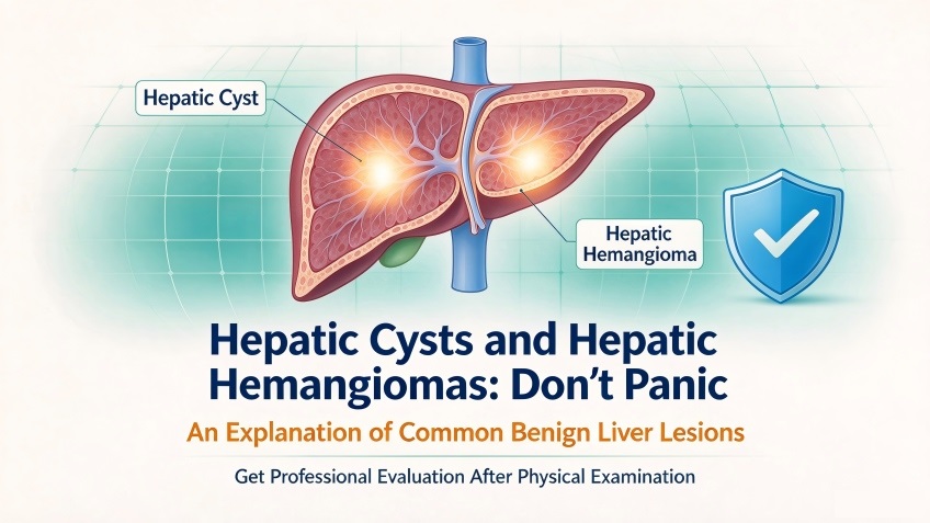
An Overview of Small, Benign Liver Lesions
In routine medical check-ups, people are sometimes surprised to find reports of abnormal liver lesions, with “hepatic cysts” and “hepatic hemangiomas” being among the most common findings. These terms can be alarming, often leading to concerns about potential malignancy. The Radiology Department at Xi’an Gaoxin Hospital provides an overview of these two benign liver conditions to help the public better understand these “minor issues.”
Hepatic Cysts: “Small Fluid-Filled Sacs” in the Liver
A hepatic cyst is a very common benign liver condition. In radiological imaging, it appears as a well-defined, homogeneous, low-density lesion—essentially a small, fluid-filled sac within the liver tissue. The sac contains clear, sterile serous fluid and has no connection to the intrahepatic bile ducts; therefore, it does not interfere with the normal secretion of bile.
Hepatic cysts are categorized as either congenital or acquired. Congenital cysts are generally believed to originate from developmental abnormalities of the intrahepatic bile ducts during the embryonic stage, while acquired cysts are often triggered by factors such as trauma, inflammation, or parasitic infections.
The vast majority of hepatic cysts cause no noticeable discomfort and are typically discovered incidentally during routine check-ups. Small, asymptomatic cysts generally require no specific treatment—periodic follow-up and observation are usually sufficient. If a cyst exceeds 5 centimeters in diameter and begins to exert pressure on surrounding organs, it may cause mild symptoms such as abdominal bloating, a dull ache in the upper right abdomen, or a loss of appetite. When it is necessary to distinguish a hepatic cyst from other liver lesions, CT or MRI scans can provide a clear diagnostic basis. A hallmark of a typical hepatic cyst on imaging is its smooth, well-defined border and uniform density; notably, it does not show “enhancement” (increased brightness) following the administration of a contrast agent—a key feature for identification.
Hepatic Hemangiomas: “Clusters of Blood Vessels” in the Liver
A hepatic hemangioma is the most common benign liver tumor, with cavernous hemangiomas accounting for the vast majority of cases. On diagnostic imaging, these lesions appear as well-defined masses within the liver, composed of a dense network of dilated, tiny blood vessels—resembling a tangled web of vessels. They are soft in texture, grow slowly, and do not invade surrounding healthy tissue.
The exact pathogenesis of hepatic hemangiomas remains not fully understood; however, the prevailing view is that they are linked to congenital vascular developmental abnormalities. Clinical research indicates that hepatic hemangiomas may tend to enlarge during periods of significant hormonal fluctuation—such as puberty or pregnancy—suggesting that hormonal changes may influence the lesions.
Most individuals with hepatic hemangiomas are asymptomatic. For lesions smaller than 5 cm that cause no discomfort, routine follow-up and observation are generally sufficient. If a lesion exceeds 5 cm in diameter, causes symptoms due to compression of adjacent structures, or is located beneath the liver capsule—posing a risk of rupture—it is necessary to seek medical attention so that a physician can determine an appropriate management plan. CT or MRI scans typically reveal characteristic enhancement patterns, which help clinicians distinguish hepatic hemangiomas from malignant liver tumors.
How to Manage a Diagnosis of Hepatic Cysts or Hepatic Hemangiomas
First and foremost, avoid excessive anxiety. Both hepatic cysts and hepatic hemangiomas are benign lesions characterized by slow growth. There is no reliable evidence to suggest that hepatic hemangiomas carry a risk of malignant transformation, and congenital hepatic cysts also grow very gradually; the vast majority of patients remain entirely asymptomatic throughout their lives.
If such lesions are detected during a medical check-up, the core principle is to follow the recommendations provided by radiologists and relevant specialists. For small lesions, the standard approach is periodic monitoring to track any changes. If a lesion meets the criteria for clinical intervention, a physician will evaluate the situation and determine the most appropriate course of action—there is no need for indiscriminate surgery or medication.