
A female patient in her 50s, who had been experiencing persistent vaginal bleeding for over a year, sought medical attention at a hospital due to sudden abdominal pain. A pelvic MRI revealed a malignant lesion in the cervix. In search of a better treatment plan, the patient and her family traveled to Guangzhou Taihe Cancer Hospital for further diagnosis and treatment.
Based on the pelvic MRI findings from the previous hospital, the initial diagnosis was highly suspected to be cervical cancer. However, after conducting a thorough physical examination, Professor Chen Jianguo, a specially invited senior expert at the hospital, and gynecologic oncologist Lin Weiwei discovered that although the patient’s cervix was significantly enlarged, the bilateral paracervical tissues were soft in consistency—a finding inconsistent with the typical biological behavior and visual characteristics of cervical cancer. They preliminarily determined that the patient’s condition was not conventional cervical cancer and that there was a possibility of misdiagnosis or missed diagnosis.
To accurately determine the cause, the team performed a diagnostic curettage on the patient and conducted a PET/CT scan along with a full suite of relevant auxiliary tests. Imaging results indicated an intrauterine mass, raising a high suspicion of endometrial malignancy. Subsequently, based on the postoperative pathology results from the diagnostic dilation and curettage and immunohistochemistry data, the uterine lesion was definitively diagnosed as endometrial adenocarcinoma. At the same time, a series of tests further revealed that the patient also had ascending colon cancer, constituting a rare case of dual primary malignancies.
According to clinical imaging data, the patient’s PET/CT scan revealed thickening of the intestinal wall in the upper ascending colon, accompanied by localized intussusception and elevated metabolic activity. Clinical data indicate that intussusception in adults is often caused by intestinal tumors; furthermore, the SUV value of this colonic lesion was as high as 19.5—far exceeding the 2.5 threshold for determining malignancy—indicating significant characteristics of a malignant lesion. Subsequently, the patient underwent pain-free gastrointestinal endoscopy, during which a massive mass was observed in the ascending colon near the hepatic flexure. Pathological biopsy results confirmed a villous-tubular adenocarcinoma, classified as a moderately differentiated adenocarcinoma.
Multidisciplinary Combined Surgery: One-Stop Radical Treatment for Two Lesions
In response to the patient’s particularly complex condition—endometrial cancer combined with ascending colon cancer—the hospital immediately activated its multidisciplinary team (MDT) consultation mechanism. Specialists from the gynaecology, radiology, gastrointestinal surgery, pathology, anaesthesia and medical oncology departments jointly discussed a treatment plan.
The expert panel concluded that both malignant tumours met the criteria for surgery and required radical resection. Although a combined operation would increase the surgical difficulty and prolong the duration of the procedure and anaesthesia, it would spare the patient the trauma of a second operation and maximise her long-term prognosis and personal benefit, making it the optimal treatment plan.
After comprehensive consideration of the patient’s condition and treatment risks, it was decided that surgical treatment would proceed first, with a subsequent adjuvant treatment plan to be formulated based on the pathological results and the patient’s recovery. Subsequently, the hospital’s gynaecological oncology and gastrointestinal surgery specialist teams collaborated to perform a combined surgery, providing the patient with a one-stop radical tumour resection.
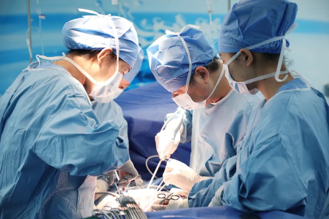
The operation was led by Professor Chen Jianguo. The gynaecological oncology team first completed a series of procedures via an abdominal approach, including a near-total hysterectomy, bilateral adnexectomy, omentectomy, pelvic lymph node dissection and para-aortic lymph node dissection. The procedure was then taken over by Dr Xu Hui, an Associate Chief Physician in the Department of Gastrointestinal Surgery, who performed a radical resection of the right half of the colon.
To minimise surgical trauma and reduce the burden on the patient’s post-operative recovery, the radical resection of the right half of the colon was performed entirely via the original gynaecological surgical approach. The position of the original incision was relatively high, which partially obstructed the surgical field of view for the colon surgery and significantly increased the difficulty of the procedure. However, to avoid extending the incision further and causing additional tissue damage, the team performed the entire procedure with precision within the existing incision, completing all surgical steps to the highest standards to ensure the best possible post-operative recovery for the patient.
The entire combined surgical procedure was successfully completed, and the patient’s post-operative condition remained stable. The patient’s family expressed high praise for the hospital’s precise diagnosis and meticulous surgical treatment, acknowledging that it was thanks to the hospital’s professional medical team that not only was the cause of the uterine lesion accurately identified, but a hidden malignant tumour in the colon was also promptly detected, enabling the patient to receive comprehensive, efficient and targeted treatment.
The incidence of multiple primary cancers is low, making them highly prone to misdiagnosis and missed diagnosis
In clinical terms, multiple primary cancers are defined as two or more independent primary malignant tumours occurring in the same patient either simultaneously or sequentially; they are primarily classified into two categories: synchronous multiple primary cancers and asynchronous multiple primary cancers. Specifically, if two or more malignant tumours of different cellular origins are detected within a six-month period, this is classified as synchronous multiple primary cancer, and the patient is considered a typical case of synchronous multiple primary cancer.
According to Dr Zhan Xiangbo, Associate Chief Physician in the Department of Gynaecological Oncology at the hospital, the clinical incidence of multiple primary cancers is extremely low; precisely for this reason, misdiagnosis is highly likely to occur in clinical practice. Once a single primary malignant tumour has been confirmed, clinicians often overlook hidden primary tumours in other sites, frequently misinterpreting multiple primary cancers as distant metastases from a single tumour, which in turn leads to deviations in the treatment plan.
Early, accurate diagnosis and standardised combined treatment of multiple primary cancers are key to ensuring effective treatment outcomes and improving survival rates. The hospital’s clinical team comprehensively reviews the patient’s medical records from other hospitals, as well as imaging and pathology reports. They conduct a precise assessment based on the patient’s individual circumstances and arrange supplementary tests as required. Always prioritising the patient’s best interests, the team develops personalised treatment plans for patients with complex tumours using precise, efficient and cost-effective diagnostic and therapeutic approaches, thereby minimising the risk of misdiagnosis or missed diagnoses and ensuring the safety and efficacy of treatment.